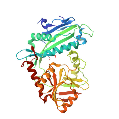
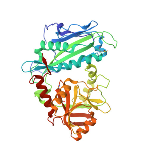
2HHF

X-ray crystal structure of oxidized human mitochondrial branched chain aminotransferase (hBCATm)


Experimental Data Snapshot

  • Method: X-RAY DIFFRACTION
  • Resolution: 1.80 Å
  • R-Value Free: 0.283 
  • R-Value Work: 0.252 
  • R-Value Observed: 0.283 

Starting Model: experimental
View more details

wwPDB Validation   3D Report Full Report


Ligand Structure Quality Assessment 


This is version 1.5 of the entry. See complete history


Literature

Human Mitochondrial Branched Chain Aminotransferase Isozyme: STRUCTURAL ROLE OF THE CXXC CENTER IN CATALYSIS.

Yennawar, N.H.Islam, M.M.Conway, M.Wallin, R.Hutson, S.M.

(2006) J Biol Chem 281: 39660-39671

  • DOI: https://doi.org/10.1074/jbc.M607552200
  • Primary Citation of Related Structures:  
    2HDK, 2HG8, 2HGW, 2HGX, 2HHF

  • PubMed Abstract: 

    Mammalian branched chain aminotransferases (BCATs) have a unique CXXC center. Kinetic and structural studies of three CXXC center mutants (C315A, C318A, and C315A/C318A) of human mitochondrial (hBCATm) isozyme and the oxidized hBCATm enzyme (hBCATm-Ox) have been used to elucidate the role of this center in hBCATm catalysis. X-ray crystallography revealed that the CXXC motif, through its network of hydrogen bonds, plays a crucial role in orienting the substrate optimally for catalysis. In all structures, there were changes in the structure of the beta-turn preceding the CXXC motif when compared with wild type protein. The N-terminal loop between residues 15 and 32 is flexible in the oxidized and mutant enzymes, the disorder greater in the oxidized protein. Disordering of the N-terminal loop disrupts the integrity of the side chain binding pocket, particularly for the branched chain side chain, less so for the dicarboxylate substrate side chain. The kinetic studies of the mutant and oxidized enzymes support the structural analysis. The kinetic results showed that the predominant effect of oxidation was on the second half-reaction rather than the first half-reaction. The oxidized enzyme was completely inactive, whereas the mutants showed limited activity. Model building of the second half-reaction substrate alpha-ketoisocaproate in the pyridoxamine 5'-phosphate-hBCATm structure suggests that disruption of the CXXC center results in altered substrate orientation and deprotonation of the amino group of pyridoxamine 5'-phosphate, which inhibits catalysis.


  • Organizational Affiliation

    Departments of Biochemistry and Internal Medicine, Wake Forest University School of Medicine, Winston-Salem, North Carolina 27157, USA.


Macromolecules
Find similar proteins by:  (by identity cutoff)  |  3D Structure
Entity ID: 1
MoleculeChains Sequence LengthOrganismDetailsImage
Branched-chain-amino-acid aminotransferase, mitochondrial365Homo sapiensMutation(s): 2 
Gene Names: BCAT2BCATMECA40
EC: 2.6.1.42
UniProt & NIH Common Fund Data Resources
Find proteins for O15382 (Homo sapiens)
Explore O15382 
Go to UniProtKB:  O15382
PHAROS:  O15382
GTEx:  ENSG00000105552 
Entity Groups  
Sequence Clusters30% Identity50% Identity70% Identity90% Identity95% Identity100% Identity
UniProt GroupO15382
Sequence Annotations
Expand
  • Reference Sequence
Find similar proteins by:  (by identity cutoff)  |  3D Structure
Entity ID: 2
MoleculeChains Sequence LengthOrganismDetailsImage
Branched-chain-amino-acid aminotransferase, mitochondrial365Homo sapiensMutation(s): 1 
Gene Names: BCAT2BCATMECA40
EC: 2.6.1.42
UniProt & NIH Common Fund Data Resources
Find proteins for O15382 (Homo sapiens)
Explore O15382 
Go to UniProtKB:  O15382
PHAROS:  O15382
GTEx:  ENSG00000105552 
Entity Groups  
Sequence Clusters30% Identity50% Identity70% Identity90% Identity95% Identity100% Identity
UniProt GroupO15382
Sequence Annotations
Expand
  • Reference Sequence
Small Molecules
Ligands 2 Unique
IDChains Name / Formula / InChI Key2D Diagram3D Interactions
PLP
Query on PLP

Download Ideal Coordinates CCD File 
C [auth A],
D [auth B]
PYRIDOXAL-5'-PHOSPHATE
C8 H10 N O6 P
NGVDGCNFYWLIFO-UHFFFAOYSA-N
EPE
Query on EPE

Download Ideal Coordinates CCD File 
E [auth B]4-(2-HYDROXYETHYL)-1-PIPERAZINE ETHANESULFONIC ACID
C8 H18 N2 O4 S
JKMHFZQWWAIEOD-UHFFFAOYSA-N
Modified Residues  2 Unique
IDChains TypeFormula2D DiagramParent
OCS
Query on OCS
A
L-PEPTIDE LINKINGC3 H7 N O5 SCYS
TYO
Query on TYO
A
L-PEPTIDE LINKINGC9 H13 N O5TYR
Experimental Data & Validation

Experimental Data

  • Method: X-RAY DIFFRACTION
  • Resolution: 1.80 Å
  • R-Value Free: 0.283 
  • R-Value Work: 0.252 
  • R-Value Observed: 0.283 
  • Space Group: P 1 21 1
Unit Cell:
Length ( Å )Angle ( ˚ )
a = 58.123α = 90
b = 104.183β = 100.72
c = 58.264γ = 90
Software Package:
Software NamePurpose
DENZOdata reduction
SCALEPACKdata scaling
CNSrefinement
PDB_EXTRACTdata extraction
ADSCdata collection
AMoREphasing

Structure Validation

View Full Validation Report


Ligand Structure Quality Assessment 


Entry History 

Deposition Data

Revision History  (Full details and data files)

  • Version 1.0: 2006-10-24
    Type: Initial release
  • Version 1.1: 2008-05-01
    Changes: Version format compliance
  • Version 1.2: 2011-07-13
    Changes: Non-polymer description, Version format compliance
  • Version 1.3: 2017-10-18
    Changes: Refinement description
  • Version 1.4: 2023-08-30
    Changes: Data collection, Database references, Derived calculations, Refinement description
  • Version 1.5: 2023-11-15
    Changes: Data collection, Derived calculations