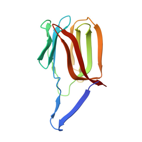
2HYR

Crystal structure of a complex of griffithsin with maltose


Experimental Data Snapshot

  • Method: X-RAY DIFFRACTION
  • Resolution: 1.51 Å
  • R-Value Free: 0.185 
  • R-Value Work: 0.152 
  • R-Value Observed: 0.154 

Starting Model: experimental
View more details

wwPDB Validation   3D Report Full Report


This is version 2.2 of the entry. See complete history


Literature

Crystallographic, thermodynamic, and molecular modeling studies of the mode of binding of oligosaccharides to the potent antiviral protein griffithsin.

Ziolkowska, N.E.Shenoy, S.R.O'keefe, B.R.McMahon, J.B.Palmer, K.E.Dwek, R.A.Wormald, M.R.Wlodawer, A.

(2007) Proteins 67: 661-670

  • DOI: https://doi.org/10.1002/prot.21336
  • Primary Citation of Related Structures:  
    2HYQ, 2HYR

  • PubMed Abstract: 

    The mode of binding of oligosaccharides to griffithsin, an antiviral lectin from the red alga Griffithsia sp., was investigated by a combination of X-ray crystallography, isothermal titration calorimetry, and molecular modeling. The structures of complexes of griffithsin with 1-->6alpha-mannobiose and with maltose were solved and refined at the resolution of 2.0 and 1.5 A, respectively. The thermodynamic parameters of binding of 1-->6alpha-mannobiose, maltose, and mannose to griffithsin were determined. Binding profiles of 1-->6alpha-mannobiose and mannose were similar with Kd values of 83.3 microM and 102 microM, respectively. The binding of maltose to griffithsin was significantly weaker, with a fourfold lower affinity (Kd = 394 microM). In all cases the binding at 30 degrees C was entropically rather than enthalpically driven. On the basis of the experimental crystal structures, as well as on previously determined structures of complexes with monosaccharides, it was possible to create a model of a tridentate complex of griffithsin with Man9GlcNAc2, a high mannose oligosaccharide commonly found on the surface of viral glycoproteins. All shorter oligomannoses could be modeled only as bidentate or monodentate complexes with griffithsin. The ability to mediate tight multivalent and multisite interactions with high-mannose oligosaccharides helps to explain the potent antiviral activity of griffithsin.


  • Organizational Affiliation

    Protein Structure Section, Macromolecular Crystallography Laboratory, National Cancer Institute, NCI-Frederick, Frederick, Maryland 21702-1201, USA.


Macromolecules
Find similar proteins by:  (by identity cutoff)  |  3D Structure
Entity ID: 1
MoleculeChains Sequence LengthOrganismDetailsImage
Griffithsin
A, B
122GriffithsiaMutation(s): 1 
UniProt
Find proteins for P84801 (Griffithsia sp. (strain Q66D336))
Explore P84801 
Go to UniProtKB:  P84801
Entity Groups  
Sequence Clusters30% Identity50% Identity70% Identity90% Identity95% Identity100% Identity
UniProt GroupP84801
Sequence Annotations
Expand
  • Reference Sequence
Oligosaccharides

Help

Entity ID: 2
MoleculeChains Length2D Diagram Glycosylation3D Interactions
alpha-D-glucopyranose-(1-4)-beta-D-glucopyranose
C, D, E, F, G
C, D, E, F, G, H
2N/A
Glycosylation Resources
GlyTouCan:  G66120GK
GlyCosmos:  G66120GK
Small Molecules
Ligands 3 Unique
IDChains Name / Formula / InChI Key2D Diagram3D Interactions
SO4
Query on SO4

Download Ideal Coordinates CCD File 
I [auth A],
J [auth A],
O [auth B]
SULFATE ION
O4 S
QAOWNCQODCNURD-UHFFFAOYSA-L
EDO
Query on EDO

Download Ideal Coordinates CCD File 
M [auth A],
N [auth A],
P [auth B]
1,2-ETHANEDIOL
C2 H6 O2
LYCAIKOWRPUZTN-UHFFFAOYSA-N
MG
Query on MG

Download Ideal Coordinates CCD File 
K [auth A],
L [auth A]
MAGNESIUM ION
Mg
JLVVSXFLKOJNIY-UHFFFAOYSA-N
Experimental Data & Validation

Experimental Data

  • Method: X-RAY DIFFRACTION
  • Resolution: 1.51 Å
  • R-Value Free: 0.185 
  • R-Value Work: 0.152 
  • R-Value Observed: 0.154 
  • Space Group: P 21 21 21
Unit Cell:
Length ( Å )Angle ( ˚ )
a = 34.82α = 90
b = 53.26β = 90
c = 118.47γ = 90
Software Package:
Software NamePurpose
REFMACrefinement
MAR345data collection
HKL-2000data reduction
DENZOdata reduction
HKL-2000data scaling
SCALEPACKdata scaling
PHASERphasing

Structure Validation

View Full Validation Report


Entry History 

Deposition Data

Revision History  (Full details and data files)

  • Version 1.0: 2007-04-24
    Type: Initial release
  • Version 1.1: 2008-05-01
    Changes: Version format compliance
  • Version 1.2: 2011-07-13
    Changes: Advisory, Version format compliance
  • Version 1.3: 2017-10-18
    Changes: Advisory, Refinement description
  • Version 2.0: 2020-07-29
    Type: Remediation
    Reason: Carbohydrate remediation
    Changes: Advisory, Atomic model, Data collection, Database references, Derived calculations, Structure summary
  • Version 2.1: 2023-08-30
    Changes: Data collection, Database references, Refinement description, Structure summary
  • Version 2.2: 2024-10-30
    Changes: Structure summary