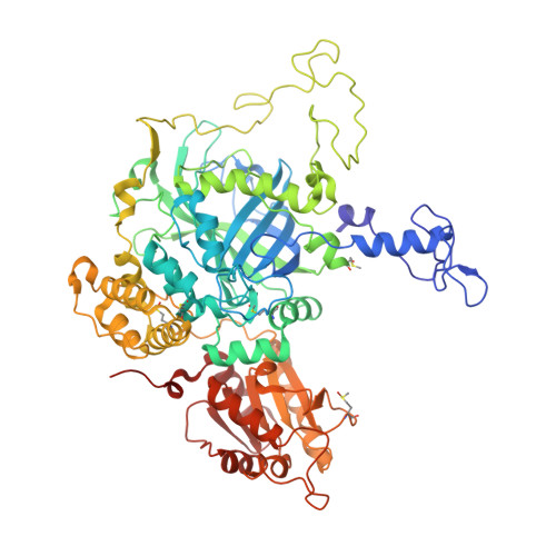
2IUF | pdb_00002iuf

The structures of Penicillium vitale catalase: resting state, oxidised state (compound I) and complex with aminotriazole


Experimental Data Snapshot

  • Method: X-RAY DIFFRACTION
  • Resolution: 1.71 Å
  • R-Value Free: 
    0.154 (Depositor), 0.162 (DCC) 
  • R-Value Work: 
    0.129 (Depositor), 0.139 (DCC) 
  • R-Value Observed: 
    0.131 (Depositor) 

wwPDB Validation   3D Report Full Report


Ligand Structure Quality Assessment 

Created with Raphaël 2.3.0Worse 01 BetterLigand structure goodness of fit to experimental dataBest fitted HDDClick on this verticalbar to view detailsBest fitted NAGClick on this verticalbar to view details

This is version 2.1 of the entry. See complete history


Literature

The Structures and Electronic Configuration of Compound I Intermediates of Helicobacter Pylori and Penicillium Vitale Catalases Determined by X-Ray Crystallography and Qm/Mm Density Functional Theory Calculations.

Alfonso-Prieto, M.Borovik, A.Carpena, X.Murshudov, G.Melik-Adamyan, W.Fita, I.Rovira, C.Loewen, P.C.

(2007) J Am Chem Soc 129: 4193

  • DOI: https://doi.org/10.1021/ja063660y
  • Primary Citation of Related Structures:  
    2IQF, 2IUF

  • PubMed Abstract: 

    The structures of Helicobacter pylori (HPC) and Penicillium vitale (PVC) catalases, each with two subunits in the crystal asymmetric unit, oxidized with peroxoacetic acid are reported at 1.8 and 1.7 A resolution, respectively. Despite the similar oxidation conditions employed, the iron-oxygen coordination length is 1.72 A for PVC, close to what is expected for a Fe=O double bond, and 1.80 and 1.85 A for HPC, suggestive of a Fe-O single bond. The structure and electronic configuration of the oxoferryl heme and immediate protein environment is investigated further by QM/MM density functional theory calculations. Four different active site electronic configurations are considered, Por*+-FeIV=O, Por*+-FeIV=O...HisH+, Por*+-FeIV-OH+ and Por-FeIV-OH (a protein radical is assumed in the latter configuration). The electronic structure of the primary oxidized species, Por*+-FeIV=O, differs qualitatively between HPC and PVC with an A2u-like porphyrin radical delocalized on the porphyrin in HPC and a mixed A1u-like "fluctuating" radical partially delocalized over the essential distal histidine, the porphyrin, and, to a lesser extent, the proximal tyrosine residue. This difference is rationalized in terms of HPC containing heme b and PVC containing heme d. It is concluded that compound I of PVC contains an oxoferryl Por*+-FeIV=O species with partial protonation of the distal histidine and compound I of HPC contains a hydroxoferryl Por-FeIV-OH with the second oxidation equivalent delocalized as a protein radical. The findings support the idea that there is a relation between radical migration to the protein and protonation of the oxoferryl bond in catalase.


  • Organizational Affiliation

    Centre especial de Recerca en Química Teorica, Parc Científic de Barcelona, Josep Samitier 1-5, 08028 Barcelona, Spain.


Macromolecules
Find similar proteins by:  (by identity cutoff)  |  3D Structure
Entity ID: 1
MoleculeChains Sequence LengthOrganismDetailsImage
CATALASEA,
B [auth E]
688Penicillium janthinellumMutation(s): 0 
EC: 1.11.1.6
UniProt
Find proteins for D9N167 (Penicillium janthinellum)
Explore D9N167 
Go to UniProtKB:  D9N167
Entity Groups  
Sequence Clusters30% Identity50% Identity70% Identity90% Identity95% Identity100% Identity
UniProt GroupD9N167
Glycosylation
Glycosylation Sites: 3
Sequence Annotations
Expand
  • Reference Sequence
Oligosaccharides

Help

Entity ID: 2
MoleculeChains Length2D Diagram Glycosylation3D Interactions
2-acetamido-2-deoxy-beta-D-glucopyranose-(1-6)-2-acetamido-2-deoxy-beta-D-glucopyranoseC [auth B]2N-Glycosylation
Glycosylation Resources
GlyTouCan:  G35932AR
GlyCosmos:  G35932AR
GlyGen:  G35932AR
Small Molecules
Ligands 7 Unique
IDChains Name / Formula / InChI Key2D Diagram3D Interactions
HDD
Query on HDD

Download Ideal Coordinates CCD File 
D [auth A],
R [auth E]
CIS-HEME D HYDROXYCHLORIN GAMMA-SPIROLACTONE
C34 H32 Fe N4 O5
UMGOPAWIGKFTRK-QQDQPIDJSA-N
NAG
Query on NAG

Download Ideal Coordinates CCD File 
E [auth A],
F [auth A],
G [auth A],
S [auth E],
T [auth E]
2-acetamido-2-deoxy-beta-D-glucopyranose
C8 H15 N O6
OVRNDRQMDRJTHS-FMDGEEDCSA-N
MPD
Query on MPD

Download Ideal Coordinates CCD File 
Q [auth A](4S)-2-METHYL-2,4-PENTANEDIOL
C6 H14 O2
SVTBMSDMJJWYQN-YFKPBYRVSA-N
F50
Query on F50

Download Ideal Coordinates CCD File 
DA [auth E],
L [auth A],
Y [auth E]
ETHANEPEROXOIC ACID
C2 H4 O3
KFSLWBXXFJQRDL-UHFFFAOYSA-N
ACT
Query on ACT

Download Ideal Coordinates CCD File 
AA [auth E]
BA [auth E]
CA [auth E]
EA [auth E]
FA [auth E]
ACETATE ION
C2 H3 O2
QTBSBXVTEAMEQO-UHFFFAOYSA-M
CA
Query on CA

Download Ideal Coordinates CCD File 
GA [auth E]
HA [auth E]
I [auth A]
J [auth A]
V [auth E]
CALCIUM ION
Ca
BHPQYMZQTOCNFJ-UHFFFAOYSA-N
O
Query on O

Download Ideal Coordinates CCD File 
H [auth A],
U [auth E]
OXYGEN ATOM
O
XLYOFNOQVPJJNP-UHFFFAOYSA-N
Modified Residues  1 Unique
IDChains TypeFormula2D DiagramParent
MHO
Query on MHO
A,
B [auth E]
L-PEPTIDE LINKINGC5 H11 N O3 SMET
Experimental Data & Validation

Experimental Data

  • Method: X-RAY DIFFRACTION
  • Resolution: 1.71 Å
  • R-Value Free:  0.154 (Depositor), 0.162 (DCC) 
  • R-Value Work:  0.129 (Depositor), 0.139 (DCC) 
  • R-Value Observed: 0.131 (Depositor) 
Space Group: P 31 2 1
Unit Cell:
Length ( Å )Angle ( ˚ )
a = 142.44α = 90
b = 142.44β = 90
c = 132.23γ = 120
Software Package:
Software NamePurpose
REFMACrefinement
DENZOdata reduction
SCALEPACKdata scaling

Structure Validation

View Full Validation Report


Ligand Structure Quality Assessment 

Created with Raphaël 2.3.0Worse 01 BetterLigand structure goodness of fit to experimental dataBest fitted HDDClick on this verticalbar to view detailsBest fitted NAGClick on this verticalbar to view details

Entry History 

Deposition Data

Revision History  (Full details and data files)

  • Version 1.0: 2006-07-10
    Type: Initial release
  • Version 1.1: 2016-12-28
    Changes: Database references, Derived calculations, Non-polymer description, Other, Structure summary, Version format compliance
  • Version 2.0: 2020-07-29
    Type: Remediation
    Reason: Carbohydrate remediation
    Changes: Advisory, Atomic model, Data collection, Derived calculations, Other, Structure summary
  • Version 2.1: 2024-11-13
    Changes: Data collection, Database references, Derived calculations, Structure summary