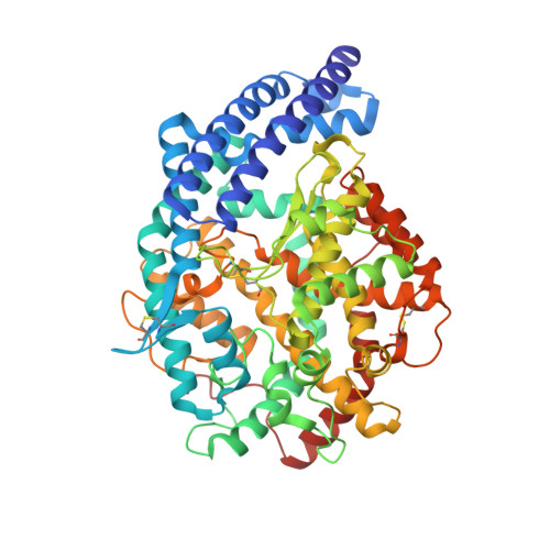
2IUL

Human tACE g13 mutant


Experimental Data Snapshot

  • Method: X-RAY DIFFRACTION
  • Resolution: 2.01 Å
  • R-Value Free: 0.220 
  • R-Value Work: 0.180 
  • R-Value Observed: 0.182 

Starting Model: experimental
View more details

wwPDB Validation   3D Report Full Report


Ligand Structure Quality Assessment 


This is version 2.2 of the entry. See complete history


Literature

Structure of Testis Ace Glycosylation Mutants and Evidence for Conserved Domain Movement.

Watermeyer, J.M.Sewell, B.T.Schwager, S.L.Natesh, R.Corradi, H.R.Acharya, K.R.Sturrock, E.D.

(2006) Biochemistry 45: 12654

  • DOI: https://doi.org/10.1021/bi061146z
  • Primary Citation of Related Structures:  
    2IUL, 2IUX

  • PubMed Abstract: 

    Human angiotensin-converting enzyme is an important drug target for which little structural information has been available until recent years. The slow progress in obtaining a crystal structure was due to the problem of surface glycosylation, a difficulty that has thus far been overcome by the use of a glucosidase-1 inhibitor in the tissue culture medium. However, the prohibitive cost of these inhibitors and incomplete glucosidase inhibition makes alternative routes to minimizing the N-glycan heterogeneity desirable. Here, glycosylation in the testis isoform (tACE) has been reduced by Asn-Gln point mutations at N-glycosylation sites, and the crystal structures of mutants having two and four intact sites have been solved to 2.0 A and 2.8 A, respectively. Both mutants show close structural identity with the wild-type. A hinge mechanism is proposed for substrate entry into the active cleft, based on homology to human ACE2 at the levels of sequence and flexibility. This is supported by normal-mode analysis that reveals intrinsic flexibility about the active site of tACE. Subdomain II, containing bound chloride and zinc ions, is found to have greater stability than subdomain I in the structures of three ACE homologues. Crystallizable glycosylation mutants open up new possibilities for cocrystallization studies to aid the design of novel ACE inhibitors.


  • Organizational Affiliation

    Division of Medical Biochemistry, Institute of Infectious Disease and Molecular Medicine, University of Cape Town, South Africa.


Macromolecules
Find similar proteins by:  (by identity cutoff)  |  3D Structure
Entity ID: 1
MoleculeChains Sequence LengthOrganismDetailsImage
ANGIOTENSIN-CONVERTING ENZYME591Homo sapiensMutation(s): 4 
EC: 3.4.15.1 (PDB Primary Data), 3.2.1 (PDB Primary Data)
UniProt & NIH Common Fund Data Resources
Find proteins for P12821 (Homo sapiens)
Explore P12821 
Go to UniProtKB:  P12821
PHAROS:  P12821
GTEx:  ENSG00000159640 
Entity Groups  
Sequence Clusters30% Identity50% Identity70% Identity90% Identity95% Identity100% Identity
UniProt GroupP12821
Glycosylation
Glycosylation Sites: 2Go to GlyGen: P12821-1
Sequence Annotations
Expand
  • Reference Sequence
Oligosaccharides

Help

Entity ID: 2
MoleculeChains Length2D Diagram Glycosylation3D Interactions
beta-D-mannopyranose-(1-3)-beta-D-mannopyranose-(1-4)-2-acetamido-2-deoxy-beta-D-glucopyranose-(1-4)-[alpha-L-fucopyranose-(1-6)]2-acetamido-2-deoxy-beta-D-glucopyranose
B
5N-Glycosylation
Glycosylation Resources
GlyTouCan:  G51347YJ
GlyCosmos:  G51347YJ
GlyGen:  G51347YJ
Small Molecules
Ligands 4 Unique
IDChains Name / Formula / InChI Key2D Diagram3D Interactions
NAG
Query on NAG

Download Ideal Coordinates CCD File 
C [auth A]2-acetamido-2-deoxy-beta-D-glucopyranose
C8 H15 N O6
OVRNDRQMDRJTHS-FMDGEEDCSA-N
ZN
Query on ZN

Download Ideal Coordinates CCD File 
H [auth A]ZINC ION
Zn
PTFCDOFLOPIGGS-UHFFFAOYSA-N
ACT
Query on ACT

Download Ideal Coordinates CCD File 
D [auth A],
E [auth A]
ACETATE ION
C2 H3 O2
QTBSBXVTEAMEQO-UHFFFAOYSA-M
CL
Query on CL

Download Ideal Coordinates CCD File 
F [auth A],
G [auth A]
CHLORIDE ION
Cl
VEXZGXHMUGYJMC-UHFFFAOYSA-M
Experimental Data & Validation

Experimental Data

  • Method: X-RAY DIFFRACTION
  • Resolution: 2.01 Å
  • R-Value Free: 0.220 
  • R-Value Work: 0.180 
  • R-Value Observed: 0.182 
  • Space Group: P 21 21 21
Unit Cell:
Length ( Å )Angle ( ˚ )
a = 56.633α = 90
b = 84.723β = 90
c = 134.466γ = 90
Software Package:
Software NamePurpose
REFMACrefinement
HKL-2000data reduction
HKL-2000data scaling

Structure Validation

View Full Validation Report


Ligand Structure Quality Assessment 


Entry History 

Deposition Data

Revision History  (Full details and data files)

  • Version 1.0: 2006-10-25
    Type: Initial release
  • Version 1.1: 2011-05-08
    Changes: Version format compliance
  • Version 1.2: 2011-07-13
    Changes: Version format compliance
  • Version 2.0: 2020-07-29
    Type: Remediation
    Reason: Carbohydrate remediation
    Changes: Advisory, Atomic model, Data collection, Derived calculations, Other, Structure summary
  • Version 2.1: 2023-12-13
    Changes: Advisory, Data collection, Database references, Refinement description, Structure summary
  • Version 2.2: 2024-11-06
    Changes: Structure summary