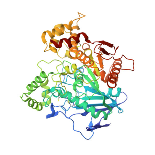
2J4C

Structure of human Butyrylcholinesterase in complex with 10mM HgCl2


Experimental Data Snapshot

  • Method: X-RAY DIFFRACTION
  • Resolution: 2.75 Å
  • R-Value Free: 0.231 
  • R-Value Work: 0.184 
  • R-Value Observed: 0.184 

Starting Model: experimental
View more details

wwPDB Validation   3D Report Full Report


Ligand Structure Quality Assessment 


This is version 3.1 of the entry. See complete history


Literature

Mechanisms of Cholinesterase Inhibition by Inorganic Mercury.

Frasco, M.F.Colletier, J.Weik, M.Carvalho, F.Guilhermino, L.Stojan, J.Fournier, D.

(2007) FEBS J 274: 1849

  • DOI: https://doi.org/10.1111/j.1742-4658.2007.05732.x
  • Primary Citation of Related Structures:  
    2J4C

  • PubMed Abstract: 

    The poorly known mechanism of inhibition of cholinesterases by inorganic mercury (HgCl2) has been studied with a view to using these enzymes as biomarkers or as biological components of biosensors to survey polluted areas. The inhibition of a variety of cholinesterases by HgCl2 was investigated by kinetic studies, X-ray crystallography, and dynamic light scattering. Our results show that when a free sensitive sulfhydryl group is present in the enzyme, as in Torpedo californica acetylcholinesterase, inhibition is irreversible and follows pseudo-first-order kinetics that are completed within 1 h in the micromolar range. When the free sulfhydryl group is not sensitive to mercury (Drosophila melanogaster acetylcholinesterase and human butyrylcholinesterase) or is otherwise absent (Electrophorus electricus acetylcholinesterase), then inhibition occurs in the millimolar range. Inhibition follows a slow binding model, with successive binding of two mercury ions to the enzyme surface. Binding of mercury ions has several consequences: reversible inhibition, enzyme denaturation, and protein aggregation, protecting the enzyme from denaturation. Mercury-induced inactivation of cholinesterases is thus a rather complex process. Our results indicate that among the various cholinesterases that we have studied, only Torpedo californica acetylcholinesterase is suitable for mercury detection using biosensors, and that a careful study of cholinesterase inhibition in a species is a prerequisite before using it as a biomarker to survey mercury in the environment.


  • Organizational Affiliation

    ICBAS, Instituto de Ciências Biomédicas de Abel Salazar, Universidade do Porto, Portugal.


Macromolecules
Find similar proteins by:  (by identity cutoff)  |  3D Structure
Entity ID: 1
MoleculeChains Sequence LengthOrganismDetailsImage
CHOLINESTERASE529Homo sapiensMutation(s): 4 
EC: 3.1.1.8
UniProt & NIH Common Fund Data Resources
Find proteins for P06276 (Homo sapiens)
Explore P06276 
Go to UniProtKB:  P06276
PHAROS:  P06276
GTEx:  ENSG00000114200 
Entity Groups  
Sequence Clusters30% Identity50% Identity70% Identity90% Identity95% Identity100% Identity
UniProt GroupP06276
Glycosylation
Glycosylation Sites: 5Go to GlyGen: P06276-1
Sequence Annotations
Expand
  • Reference Sequence
Oligosaccharides

Help

Entity ID: 2
MoleculeChains Length2D Diagram Glycosylation3D Interactions
2-acetamido-2-deoxy-beta-D-glucopyranose-(1-4)-[alpha-L-fucopyranose-(1-6)]2-acetamido-2-deoxy-beta-D-glucopyranose
B, C
3N-Glycosylation
Glycosylation Resources
GlyTouCan:  G21290RB
GlyCosmos:  G21290RB
GlyGen:  G21290RB
Small Molecules
Ligands 7 Unique
IDChains Name / Formula / InChI Key2D Diagram3D Interactions
NAG
Query on NAG

Download Ideal Coordinates CCD File 
D [auth A],
E [auth A],
F [auth A]
2-acetamido-2-deoxy-beta-D-glucopyranose
C8 H15 N O6
OVRNDRQMDRJTHS-FMDGEEDCSA-N
HG
Query on HG

Download Ideal Coordinates CCD File 
H [auth A],
I [auth A],
J [auth A],
T [auth A]
MERCURY (II) ION
Hg
BQPIGGFYSBELGY-UHFFFAOYSA-N
MES
Query on MES

Download Ideal Coordinates CCD File 
P [auth A]2-(N-MORPHOLINO)-ETHANESULFONIC ACID
C6 H13 N O4 S
SXGZJKUKBWWHRA-UHFFFAOYSA-N
SO4
Query on SO4

Download Ideal Coordinates CCD File 
K [auth A],
Q [auth A]
SULFATE ION
O4 S
QAOWNCQODCNURD-UHFFFAOYSA-L
GOL
Query on GOL

Download Ideal Coordinates CCD File 
L [auth A],
M [auth A],
N [auth A]
GLYCEROL
C3 H8 O3
PEDCQBHIVMGVHV-UHFFFAOYSA-N
BUA
Query on BUA

Download Ideal Coordinates CCD File 
O [auth A]butanoic acid
C4 H8 O2
FERIUCNNQQJTOY-UHFFFAOYSA-N
CL
Query on CL

Download Ideal Coordinates CCD File 
G [auth A],
R [auth A],
S [auth A]
CHLORIDE ION
Cl
VEXZGXHMUGYJMC-UHFFFAOYSA-M
Modified Residues  1 Unique
IDChains TypeFormula2D DiagramParent
CSS
Query on CSS
A
L-PEPTIDE LINKINGC3 H7 N O2 S2CYS
Experimental Data & Validation

Experimental Data

  • Method: X-RAY DIFFRACTION
  • Resolution: 2.75 Å
  • R-Value Free: 0.231 
  • R-Value Work: 0.184 
  • R-Value Observed: 0.184 
  • Space Group: I 4 2 2
Unit Cell:
Length ( Å )Angle ( ˚ )
a = 153.76α = 90
b = 153.76β = 90
c = 128.58γ = 90
Software Package:
Software NamePurpose
CNSrefinement
XDSdata reduction
XSCALEdata scaling
CNSphasing

Structure Validation

View Full Validation Report


Ligand Structure Quality Assessment 


Entry History 

Deposition Data

Revision History  (Full details and data files)

  • Version 1.0: 2007-03-27
    Type: Initial release
  • Version 1.1: 2011-05-08
    Changes: Version format compliance
  • Version 1.2: 2011-07-13
    Changes: Version format compliance
  • Version 2.0: 2019-01-30
    Changes: Atomic model, Data collection, Experimental preparation
  • Version 2.1: 2019-02-06
    Changes: Data collection, Experimental preparation
  • Version 3.0: 2020-07-29
    Type: Remediation
    Reason: Carbohydrate remediation
    Changes: Atomic model, Data collection, Derived calculations, Other, Structure summary
  • Version 3.1: 2023-12-13
    Changes: Data collection, Database references, Derived calculations, Refinement description, Structure summary