Crystal Structure of Human Inosine Triphosphatase: Substrate Binding and Implication of the Inosine Triphosphatase Deficiency Mutation P32T.
Stenmark, P., Kursula, P., Flodin, S., Graslund, S., Landry, R., Nordlund, P., Schuler, H.(2007) J Biol Chem 282: 3182
- PubMed: 17138556
- DOI: https://doi.org/10.1074/jbc.M609838200
- Primary Citation of Related Structures:
2CAR, 2J4E - PubMed Abstract:
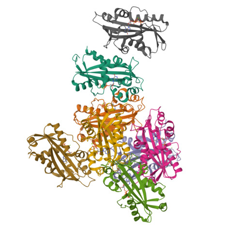
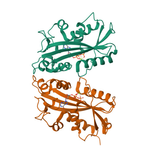
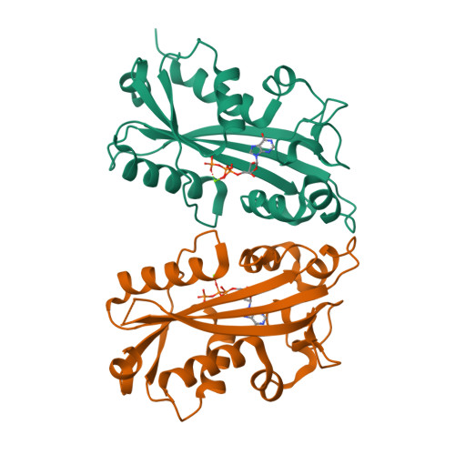
Inosine triphosphatase (ITPA) is a ubiquitous key regulator of cellular non-canonical nucleotide levels. It breaks down inosine and xanthine nucleotides generated by deamination of purine bases. Its enzymatic action prevents accumulation of ITP and reduces the risk of incorporation of potentially mutagenic inosine nucleotides into nucleic acids. Here we describe the crystal structure of human ITPA in complex with its prime substrate ITP, as well as the apoenzyme at 2.8 and 1.1A, respectively. These structures show for the first time the site of substrate and Mg2+ coordination as well as the conformational changes accompanying substrate binding in this class of enzymes. Enzyme substrate interactions induce an extensive closure of the nucleotide binding grove, resulting in tight interactions with the base that explain the high substrate specificity of ITPA for inosine and xanthine over the canonical nucleotides. One of the dimer contact sites is made up by a loop that is involved in coordinating the metal ion in the active site. We predict that the ITPA deficiency mutation P32T leads to a shift of this loop that results in a disturbed affinity for nucleotides and/or a reduced catalytic activity in both monomers of the physiological dimer.
Organizational Affiliation:
Structural Genomics Consortium, Department of Medical Biochemistry and Biophysics, Karolinska Institute, 17177 Stockholm, Sweden.


