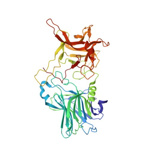
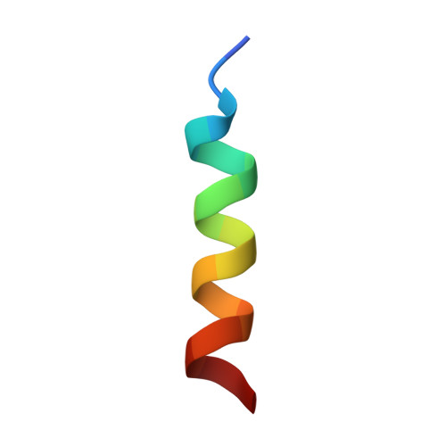
2NM1

Structure of BoNT/B in complex with its protein receptor


Experimental Data Snapshot

  • Method: X-RAY DIFFRACTION
  • Resolution: 2.15 Å
  • R-Value Free: 0.246 
  • R-Value Work: 0.199 
  • R-Value Observed: 0.199 

Starting Model: experimental
View more details

wwPDB Validation   3D Report Full Report


This is version 1.3 of the entry. See complete history


Literature

Botulinum neurotoxin B recognizes its protein receptor with high affinity and specificity.

Jin, R.Rummel, A.Binz, T.Brunger, A.T.

(2006) Nature 444: 1092-1095

  • DOI: https://doi.org/10.1038/nature05387
  • Primary Citation of Related Structures:  
    2NM1

  • PubMed Abstract: 

    Botulinum neurotoxins (BoNTs) are produced by Clostridium botulinum and cause the neuroparalytic syndrome of botulism. With a lethal dose of 1 ng kg(-1), they pose a biological hazard to humans and a serious potential bioweapon threat. BoNTs bind with high specificity at neuromuscular junctions and they impair exocytosis of synaptic vesicles containing acetylcholine through specific proteolysis of SNAREs (soluble N-ethylmaleimide-sensitive fusion protein attachment protein receptors), which constitute part of the synaptic vesicle fusion machinery. The molecular details of the toxin-cell recognition have been elusive. Here we report the structure of a BoNT in complex with its protein receptor: the receptor-binding domain of botulinum neurotoxin serotype B (BoNT/B) bound to the luminal domain of synaptotagmin II, determined at 2.15 A resolution. On binding, a helix is induced in the luminal domain which binds to a saddle-shaped crevice on a distal tip of BoNT/B. This crevice is adjacent to the non-overlapping ganglioside-binding site of BoNT/B. Synaptotagmin II interacts with BoNT/B with nanomolar affinity, at both neutral and acidic endosomal pH. Biochemical and neuronal ex vivo studies of structure-based mutations indicate high specificity and affinity of the interaction, and high selectivity of BoNT/B among synaptotagmin I and II isoforms. Synergistic binding of both synaptotagmin and ganglioside imposes geometric restrictions on the initiation of BoNT/B translocation after endocytosis. Our results provide the basis for the rational development of preventive vaccines or inhibitors against these neurotoxins.


  • Organizational Affiliation

    Howard Hughes Medical Institute, Stanford University, Stanford, California 94305, USA.


Macromolecules
Find similar proteins by:  (by identity cutoff)  |  3D Structure
Entity ID: 1
MoleculeChains Sequence LengthOrganismDetailsImage
Botulinum neurotoxin type B436Clostridium botulinumMutation(s): 0 
Gene Names: botB
EC: 3.4.24.69
UniProt
Find proteins for P10844 (Clostridium botulinum)
Explore P10844 
Go to UniProtKB:  P10844
Entity Groups  
Sequence Clusters30% Identity50% Identity70% Identity90% Identity95% Identity100% Identity
UniProt GroupP10844
Sequence Annotations
Expand
  • Reference Sequence

Find similar proteins by:  Sequence   |   3D Structure  

Entity ID: 2
MoleculeChains Sequence LengthOrganismDetailsImage
Synaptotagmin-217Rattus norvegicusMutation(s): 0 
UniProt
Find proteins for P29101 (Rattus norvegicus)
Explore P29101 
Go to UniProtKB:  P29101
Entity Groups  
Sequence Clusters30% Identity50% Identity70% Identity90% Identity95% Identity100% Identity
UniProt GroupP29101
Sequence Annotations
Expand
  • Reference Sequence
Experimental Data & Validation

Experimental Data

  • Method: X-RAY DIFFRACTION
  • Resolution: 2.15 Å
  • R-Value Free: 0.246 
  • R-Value Work: 0.199 
  • R-Value Observed: 0.199 
  • Space Group: P 21 21 21
Unit Cell:
Length ( Å )Angle ( ˚ )
a = 55.82α = 90
b = 97.52β = 90
c = 113.57γ = 90
Software Package:
Software NamePurpose
CNSrefinement
ADSCdata collection
HKL-2000data reduction
HKL-2000data scaling
PHASERphasing

Structure Validation

View Full Validation Report


Entry History 

Deposition Data

Revision History  (Full details and data files)

  • Version 1.0: 2006-12-19
    Type: Initial release
  • Version 1.1: 2008-05-01
    Changes: Version format compliance
  • Version 1.2: 2011-07-13
    Changes: Version format compliance
  • Version 1.3: 2023-08-30
    Changes: Data collection, Database references, Refinement description