2PR9 | pdb_00002pr9

Mu2 adaptin subunit (AP50) of AP2 adaptor (second domain), complexed with GABAA receptor-gamma2 subunit-derived internalization peptide DEEYGYECL


Experimental Data Snapshot

  • Method: X-RAY DIFFRACTION
  • Resolution: 2.51 Å
  • R-Value Free: 
    0.240 (Depositor), 0.246 (DCC) 
  • R-Value Work: 
    0.203 (Depositor) 
  • R-Value Observed: 
    0.204 (Depositor) 

Starting Model: experimental
View more details

wwPDB Validation 3D Report Full Report

Validation slider image for 2PR9

This is version 1.2 of the entry. See complete history

Literature

Regulation of synaptic inhibition by phospho-dependent binding of the AP2 complex to a YECL motif in the GABAA receptor gamma2 subunit.

Kittler, J.T.Chen, G.Kukhtina, V.Vahedi-Faridi, A.Gu, Z.Tretter, V.Smith, K.R.McAinsh, K.Arancibia-Carcamo, I.L.Saenger, W.Haucke, V.Yan, Z.Moss, S.J.

(2008) Proc Natl Acad Sci U S A 105: 3616-3621

  • DOI: https://doi.org/10.1073/pnas.0707920105
  • Primary Citation Related Structures: 
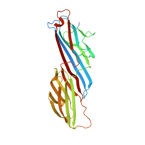
    2PR9

  • PubMed Abstract: 

    The regulation of the number of gamma2-subunit-containing GABA(A) receptors (GABA(A)Rs) present at synapses is critical for correct synaptic inhibition and animal behavior. This regulation occurs, in part, by the controlled removal of receptors from the membrane in clathrin-coated vesicles, but it remains unclear how clathrin recruitment to surface gamma2-subunit-containing GABA(A)Rs is regulated. Here, we identify a gamma2-subunit-specific Yxxvarphi-type-binding motif for the clathrin adaptor protein, AP2, which is located within a site for gamma2-subunit tyrosine phosphorylation. Blocking GABA(A)R-AP2 interactions via this motif increases synaptic responses within minutes. Crystallographic and biochemical studies reveal that phosphorylation of the Yxxvarphi motif inhibits AP2 binding, leading to increased surface receptor number. In addition, the crystal structure provides an explanation for the high affinity of this motif for AP2 and suggests that gamma2-subunit-containing heteromeric GABA(A)Rs may be internalized as dimers or multimers. These data define a mechanism for tyrosine kinase regulation of GABA(A)R surface levels and synaptic inhibition.


  • Organizational Affiliation
    • Department of Physiology, University College London, Gower Street, London WC1E 6BT, United Kingdom. j.kittler@ucl.ac.uk

Macromolecule Content 

  • Total Structure Weight: 35.34 kDa 
  • Atom Count: 2,174 
  • Modeled Residue Count: 264 
  • Deposited Residue Count: 309 
  • Unique protein chains: 2

Macromolecules

Find similar proteins by:|  3D Structure
Entity ID: 1
MoleculeChains  Sequence LengthOrganismDetailsImage
AP-2 complex subunit mu-1299Rattus norvegicusMutation(s): 0 
Gene Names: Ap2m1
UniProt
Find proteins for P84092 (Rattus norvegicus)
Explore P84092 
Go to UniProtKB:  P84092
Entity Groups
Sequence Clusters30% Identity50% Identity70% Identity90% Identity95% Identity100% Identity
UniProt GroupP84092
Sequence Annotations
Expand
Reference Sequence
Find similar proteins by:  Sequence   |   3D Structure  
Entity ID: 2
MoleculeChains  Sequence LengthOrganismDetailsImage
GABA(A) receptor subunit gamma-2 peptideB [auth P]10N/AMutation(s): 0 
UniProt
Find proteins for P18508 (Rattus norvegicus)
Explore P18508 
Go to UniProtKB:  P18508
Entity Groups
Sequence Clusters30% Identity50% Identity70% Identity90% Identity95% Identity100% Identity
UniProt GroupP18508
Sequence Annotations
Expand
Reference Sequence

Experimental Data & Validation

Experimental Data

  • Method: X-RAY DIFFRACTION
  • Resolution: 2.51 Å
  • R-Value Free:  0.240 (Depositor), 0.246 (DCC) 
  • R-Value Work:  0.203 (Depositor) 
  • R-Value Observed: 0.204 (Depositor) 
Space Group: P 64
Unit Cell:
Length ( Å )Angle ( ˚ )
a = 126.3α = 90
b = 126.3β = 90
c = 74.65γ = 120
Software Package:
Software NamePurpose
XSCALEdata scaling
MOLREPphasing
REFMACrefinement
PDB_EXTRACTdata extraction
ADSCdata collection
XDSdata reduction

Structure Validation

View Full Validation Report


Entry History 

Revision History  (Full details and data files)

  • Version 1.0: 2008-03-18
    Type: Initial release
  • Version 1.1: 2011-07-13
    Changes: Advisory, Version format compliance
  • Version 1.2: 2023-08-30
    Changes: Data collection, Database references, Refinement description