Refined structure of porcine pepsinogen at 1.8 A resolution.
Sielecki, A.R., Fujinaga, M., Read, R.J., James, M.N.(1991) J Mol Biology 219: 671-692
- PubMed: 2056534 Search on PubMed
- DOI: https://doi.org/10.1016/0022-2836(91)90664-r
- Primary Citation Related Structures:
2PSG - PubMed Abstract:
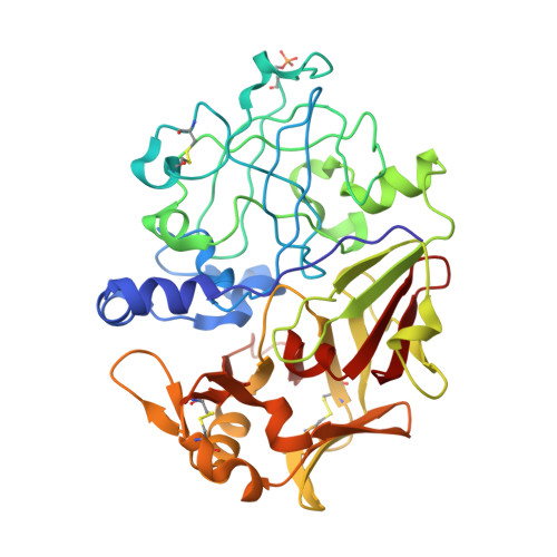
The molecular structure of porcine pepsinogen at 1.8 A resolution has been determined by a combination of molecular replacement and multiple isomorphous phasing techniques. The resulting structure was refined by restrained-parameter least-squares methods. The final R factor [formula: see text] is 0.164 for 32,264 reflections with I greater than or equal to sigma (I) in the resolution range of 8.0 to 1.8 A. The model consists of 2785 protein atoms in 370 residues, a phosphoryl group on Ser68 and 238 ordered water molecules. The resulting molecular stereochemistry is consistent with a well-refined crystal structure with co-ordinate accuracy in the range of 0.10 to 0.15 A for the well-ordered regions of the molecule (B less than 15 A2). For the enzyme portion of the zymogen, the root-mean-square difference in C alpha atom co-ordinates with the refined porcine pepsin structure is 0.90 A (284 common atoms) and with the C alpha atoms of penicillopepsin it is 1.63 A (275 common atoms). The additional 44 N-terminal amino acids of the prosegment (Leu1p to Leu44p, using the letter p after the residue number to distinguish the residues of the prosegment) adopt a relatively compact structure consisting of a long beta-strand followed by two approximately orthogonal alpha-helices and a short 3(10)-helix. Intimate contacts, both electrostatic and hydrophobic interactions, are made with residues in the pepsin active site. The N-terminal beta-strand, Leu1p to Leu6p, forms part of the six-stranded beta-sheet common to the aspartic proteinases. In the zymogen the first 13 residues of pepsin, Ile1 to Glu13, adopt a completely different conformation from that of the mature enzyme. The C alpha atom of Ile1 must move approximately 44 A in going from its position in the inactive zymogen to its observed position in active pepsin. Electrostatic interactions of Lys36pN and hydrogen-bonding interactions of Tyr37pOH, and Tyr90H with the two catalytic aspartate groups, Asp32 and Asp215, prevent substrate access to the active site of the zymogen. We have made a detailed comparison of the mammalian pepsinogen fold with the fungal aspartic proteinase fold of penicillopepsin, used for the molecular replacement solution. A structurally derived alignment of the two sequences is presented.
- Department of Biochemistry, University of Alberta, Edmonton, Canada.
Organizational Affiliation: