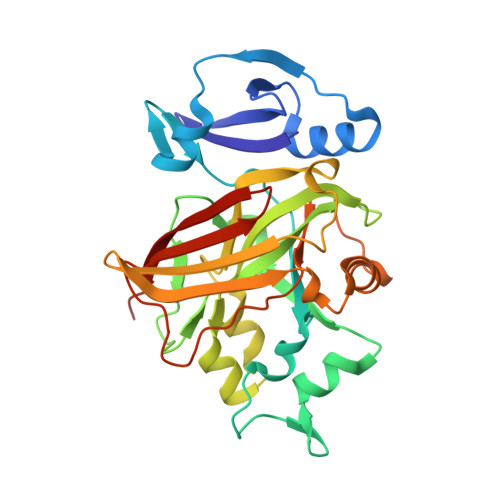
2Q1A

2-keto-3-deoxy-D-arabinonate dehydratase complexed with magnesium and 2-oxobutyrate


Experimental Data Snapshot

  • Method: X-RAY DIFFRACTION
  • Resolution: 2.50 Å
  • R-Value Free: 0.256 
  • R-Value Work: 0.234 
  • R-Value Observed: 0.235 

Starting Model: experimental
View more details

wwPDB Validation   3D Report Full Report


This is version 2.0 of the entry. See complete history


Literature

Structural insight into substrate binding and catalysis of a novel 2-keto-3-deoxy-D-arabinonate dehydratase illustrates common mechanistic features of the FAH superfamily.

Brouns, S.J.Barends, T.R.Worm, P.Akerboom, J.Turnbull, A.P.Salmon, L.van der Oost, J.

(2008) J Mol Biol 379: 357-371

  • DOI: https://doi.org/10.1016/j.jmb.2008.03.064
  • Primary Citation of Related Structures:  
    2Q18, 2Q19, 2Q1A, 2Q1C, 2Q1D, 3BQB

  • PubMed Abstract: 

    The archaeon Sulfolobus solfataricus converts d-arabinose to 2-oxoglutarate by an enzyme set consisting of two dehydrogenases and two dehydratases. The third step of the pathway is catalyzed by a novel 2-keto-3-deoxy-D-arabinonate dehydratase (KdaD). In this study, the crystal structure of the enzyme has been solved to 2.1 A resolution. The enzyme forms an oval-shaped ring of four subunits, each consisting of an N-terminal domain with a four-stranded beta-sheet flanked by two alpha-helices, and a C-terminal catalytic domain with a fumarylacetoacetate hydrolase (FAH) fold. Crystal structures of complexes of the enzyme with magnesium or calcium ions and either a substrate analog 2-oxobutyrate, or the aldehyde enzyme product 2,5-dioxopentanoate revealed that the divalent metal ion in the active site is coordinated octahedrally by three conserved carboxylate residues, a water molecule, and both the carboxylate and the oxo groups of the substrate molecule. An enzymatic mechanism for base-catalyzed dehydration is proposed on the basis of the binding mode of the substrate to the metal ion, which suggests that the enzyme enhances the acidity of the protons alpha to the carbonyl group, facilitating their abstraction by glutamate 114. A comprehensive structural comparison of members of the FAH superfamily is presented and their evolution is discussed, providing a basis for functional investigations of this largely unexplored protein superfamily.


  • Organizational Affiliation

    Laboratory of Microbiology, Department of Agrotechnology and Food Sciences, Wageningen University, Dreienplein 10, 6703 HB Wageningen, Netherlands. stan.brouns@wur.nl


Macromolecules
Find similar proteins by:  (by identity cutoff)  |  3D Structure
Entity ID: 1
MoleculeChains Sequence LengthOrganismDetailsImage
2-keto-3-deoxy-D-arabinonate dehydrataseA [auth X]293Saccharolobus solfataricus P2Mutation(s): 0 
Gene Names: kdaD
EC: 4.2.1.141
UniProt
Find proteins for Q97UA0 (Saccharolobus solfataricus (strain ATCC 35092 / DSM 1617 / JCM 11322 / P2))
Explore Q97UA0 
Go to UniProtKB:  Q97UA0
Entity Groups  
Sequence Clusters30% Identity50% Identity70% Identity90% Identity95% Identity100% Identity
UniProt GroupQ97UA0
Sequence Annotations
Expand
  • Reference Sequence
Small Molecules
Ligands 2 Unique
IDChains Name / Formula / InChI Key2D Diagram3D Interactions
2KT
Query on 2KT

Download Ideal Coordinates CCD File 
C [auth X]2-KETOBUTYRIC ACID
C4 H6 O3
TYEYBOSBBBHJIV-UHFFFAOYSA-N
MG
Query on MG

Download Ideal Coordinates CCD File 
B [auth X]MAGNESIUM ION
Mg
JLVVSXFLKOJNIY-UHFFFAOYSA-N
Experimental Data & Validation

Experimental Data

  • Method: X-RAY DIFFRACTION
  • Resolution: 2.50 Å
  • R-Value Free: 0.256 
  • R-Value Work: 0.234 
  • R-Value Observed: 0.235 
  • Space Group: I 41 2 2
Unit Cell:
Length ( Å )Angle ( ˚ )
a = 128.83α = 90
b = 128.83β = 90
c = 224.4γ = 90
Software Package:
Software NamePurpose
REFMACrefinement
MAR345dtbdata collection
XDSdata reduction
XSCALEdata scaling
XFITdata reduction

Structure Validation

View Full Validation Report


Entry History 

Deposition Data

Revision History  (Full details and data files)

  • Version 1.0: 2008-04-08
    Type: Initial release
  • Version 1.1: 2011-07-13
    Changes: Source and taxonomy, Version format compliance
  • Version 1.2: 2023-08-30
    Changes: Data collection, Database references, Derived calculations, Refinement description
  • Version 2.0: 2023-11-15
    Changes: Atomic model, Data collection, Derived calculations