2QQ0

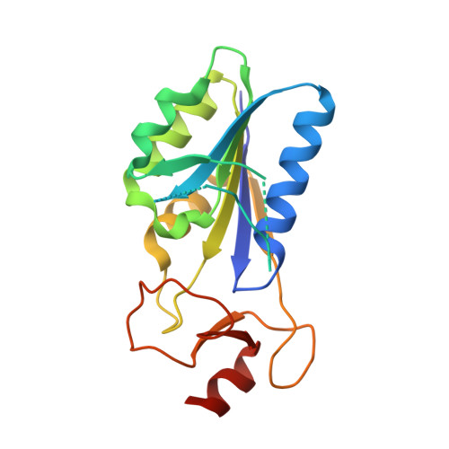
Thymidine Kinase from Thermotoga Maritima in complex with thymidine + AppNHp


Experimental Data Snapshot

  • Method: X-RAY DIFFRACTION
  • Resolution: 1.50 Å
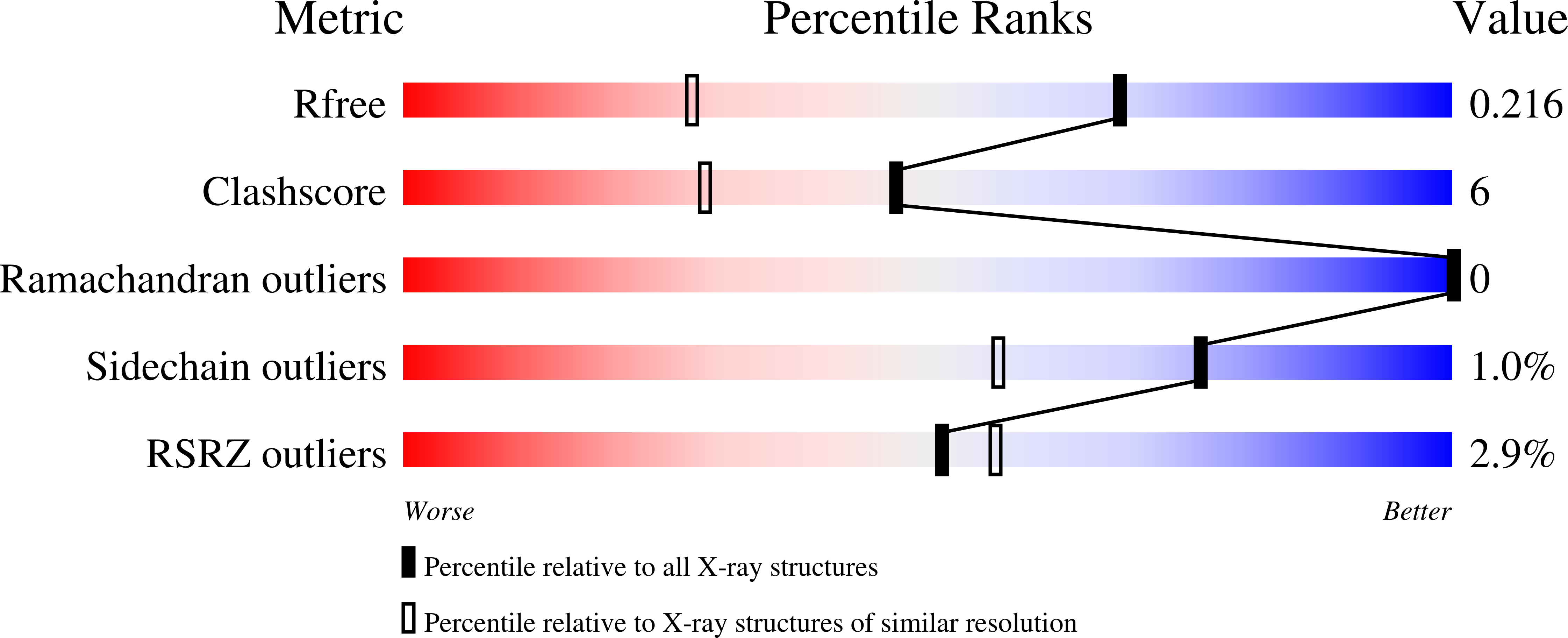
  • R-Value Free: 0.217 
  • R-Value Work: 0.171 
  • R-Value Observed: 0.175 

wwPDB Validation   3D Report Full Report


Ligand Structure Quality Assessment 


This is version 1.2 of the entry. See complete history


Literature

Quaternary structure change as a mechanism for the regulation of thymidine kinase 1-like enzymes.

Segura-Pena, D.Lichter, J.Trani, M.Konrad, M.Lavie, A.Lutz, S.

(2007) Structure 15: 1555-1566

  • DOI: https://doi.org/10.1016/j.str.2007.09.025
  • Primary Citation of Related Structures:  
    2QPO, 2QQ0, 2QQE

  • PubMed Abstract: 

    The human cytosolic thymidine kinase (TK) and structurally related TKs in prokaryotes play a crucial role in the synthesis and regulation of the cellular thymidine triphosphate pool. We report the crystal structures of the TK homotetramer from Thermotoga maritima in four different states: its apo-form, a binary complex with thymidine, as well as the ternary structures with the two substrates (thymidine/AppNHp) and the reaction products (TMP/ADP). In combination with fluorescence spectroscopy and mutagenesis experiments, our results demonstrate that ATP binding is linked to a substantial reorganization of the enzyme quaternary structure, leading to a transition from a closed, inactive conformation to an open, catalytic state. We hypothesize that these structural changes are relevant to enzyme function in situ as part of the catalytic cycle and serve an important role in regulating enzyme activity by amplifying the effects of feedback inhibitor binding.


  • Organizational Affiliation

    Department of Biochemistry and Molecular Genetics, University of Illinois at Chicago, 900 South Ashland Avenue, Chicago, IL 60607, USA.


Macromolecules
Find similar proteins by:  (by identity cutoff)  |  3D Structure
Entity ID: 1
MoleculeChains Sequence LengthOrganismDetailsImage
Thymidine kinase
A, B
184Thermotoga maritimaMutation(s): 0 
Gene Names: tdk
EC: 2.7.1.21
UniProt
Find proteins for Q9WYN2 (Thermotoga maritima (strain ATCC 43589 / DSM 3109 / JCM 10099 / NBRC 100826 / MSB8))
Go to UniProtKB:  Q9WYN2
Sequence Annotations
Expand
  • Reference Sequence
Small Molecules
Ligands 6 Unique
IDChains Name / Formula / InChI Key2D Diagram3D Interactions
ANP
Query on ANP

Download Ideal Coordinates CCD File 
F [auth A]PHOSPHOAMINOPHOSPHONIC ACID-ADENYLATE ESTER
C10 H17 N6 O12 P3
PVKSNHVPLWYQGJ-KQYNXXCUSA-N
ADP
Query on ADP

Download Ideal Coordinates CCD File 
J [auth B]ADENOSINE-5'-DIPHOSPHATE
C10 H15 N5 O10 P2
XTWYTFMLZFPYCI-KQYNXXCUSA-N
TMP
Query on TMP

Download Ideal Coordinates CCD File 
I [auth B]THYMIDINE-5'-PHOSPHATE
C10 H15 N2 O8 P
GYOZYWVXFNDGLU-XLPZGREQSA-N
THM
Query on THM

Download Ideal Coordinates CCD File 
E [auth A]THYMIDINE
C10 H14 N2 O5
IQFYYKKMVGJFEH-XLPZGREQSA-N
ZN
Query on ZN

Download Ideal Coordinates CCD File 
C [auth A],
G [auth B]
ZINC ION
Zn
PTFCDOFLOPIGGS-UHFFFAOYSA-N
MG
Query on MG

Download Ideal Coordinates CCD File 
D [auth A],
H [auth B]
MAGNESIUM ION
Mg
JLVVSXFLKOJNIY-UHFFFAOYSA-N
Experimental Data & Validation

Experimental Data

  • Method: X-RAY DIFFRACTION
  • Resolution: 1.50 Å
  • R-Value Free: 0.217 
  • R-Value Work: 0.171 
  • R-Value Observed: 0.175 
  • Space Group: C 1 2 1
Unit Cell:
Length ( Å )Angle ( ˚ )
a = 102.16α = 90
b = 59.31β = 103.02
c = 61.3γ = 90
Software Package:
Software NamePurpose
REFMACrefinement
XDSdata scaling
XDSdata reduction
MOLREPphasing

Structure Validation

View Full Validation Report


Ligand Structure Quality Assessment 


Entry History 

Deposition Data

Revision History  (Full details and data files)

  • Version 1.0: 2007-10-16
    Type: Initial release
  • Version 1.1: 2011-07-13
    Changes: Version format compliance
  • Version 1.2: 2024-02-21
    Changes: Data collection, Database references, Derived calculations