2V6A

Crystal structure of Chlamydomonas reinhardtii Rubisco with large- subunit mutations V331A, G344S


Experimental Data Snapshot

  • Method: X-RAY DIFFRACTION
  • Resolution: 1.50 Å
  • R-Value Free: 
    0.192 (Depositor), 0.180 (DCC) 
  • R-Value Work: 
    0.176 (Depositor), 0.180 (DCC) 
  • R-Value Observed: 
    0.177 (Depositor) 

Starting Model: experimental
View more details

wwPDB Validation   3D Report Full Report


Ligand Structure Quality Assessment 

Created with Raphaël 2.3.0Worse 01 BetterLigand structure goodness of fit to experimental dataBest fitted CAPClick on this verticalbar to view details

This is version 1.3 of the entry. See complete history


Literature

Structural Analysis of Altered Large-Subunit Loop-6-Carboxy-Terminus Interactions that Influence Catalytic Efficiency and Co2-O2 Specificity of Ribulose-1,5-Bisphosphate Carboxylase Oxygenase

Karkehabadi, S.Satagopan, S.Taylor, T.C.Spreitzer, R.J.Andersson, I.

(2007) Biochemistry 46: 11080

  • DOI: https://doi.org/10.1021/bi701063f
  • Primary Citation of Related Structures:  
    2V63, 2V67, 2V68, 2V69, 2V6A

  • PubMed Abstract: 

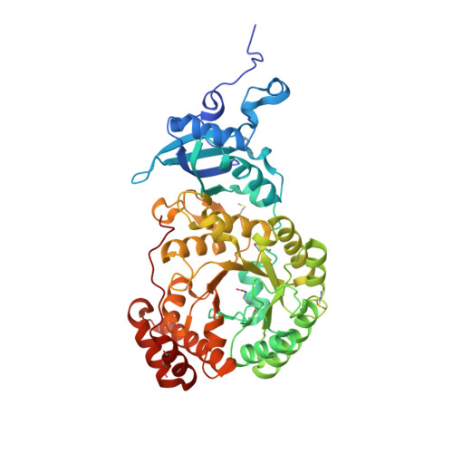
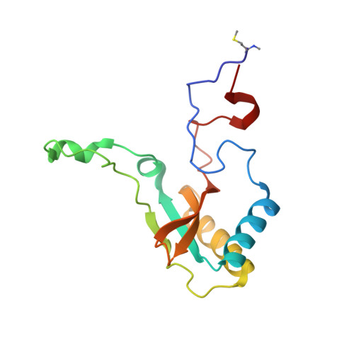
    The loop between alpha-helix 6 and beta-strand 6 in the alpha/beta-barrel of ribulose-1,5-bisphosphate carboxylase/oxygenase plays a key role in discriminating between CO2 and O2. Genetic screening in Chlamydomonas reinhardtii previously identified a loop-6 V331A substitution that decreases carboxylation and CO2/O2 specificity. Revertant selection identified T342I and G344S substitutions that restore photosynthetic growth by increasing carboxylation and specificity of the V331A enzyme. In numerous X-ray crystal structures, loop 6 is closed or open depending on the activation state of the enzyme and the presence or absence of ligands. The carboxy terminus folds over loop 6 in the closed state. To study the molecular basis for catalysis, directed mutagenesis and chloroplast transformation were used to create T342I and G344S substitutions alone. X-ray crystal structures were then solved for the V331A, V331A/T342I, T342I, and V331A/G344S enzymes, as well as for a D473E enzyme created to assess the role of the carboxy terminus in loop-6 closure. V331A disturbs a hydrophobic pocket, abolishing several van der Waals interactions. These changes are complemented by T342I and G344S, both of which alone cause decreases in CO2/O2 specificity. In the V331A/T342I revertant enzyme, Arg339 main-chain atoms are displaced. In V331A/G344S, alpha-helix 6 is shifted. D473E causes disorder of the carboxy terminus, but loop 6 remains closed. Interactions between a transition-state analogue and several residues are altered in the mutant enzymes. However, active-site Lys334 at the apex of loop 6 has a normal conformation. A variety of subtle interactions must be responsible for catalytic efficiency and CO2/O2 specificity.


  • Organizational Affiliation

    Department of Molecular Biology, Swedish University of Agricultural Sciences, BMC Box 590, 751 24 Uppsala, Sweden.


Macromolecules
Find similar proteins by:  (by identity cutoff)  |  3D Structure
Entity ID: 1
MoleculeChains Sequence LengthOrganismDetailsImage
RIBULOSE BISPHOSPHATE CARBOXYLASE LARGE CHAIN
A, B, C, D, E
475Chlamydomonas reinhardtiiMutation(s): 2 
EC: 4.1.1.39
UniProt
Find proteins for P00877 (Chlamydomonas reinhardtii)
Explore P00877 
Go to UniProtKB:  P00877
Entity Groups  
Sequence Clusters30% Identity50% Identity70% Identity90% Identity95% Identity100% Identity
UniProt GroupP00877
Sequence Annotations
Expand
  • Reference Sequence
Find similar proteins by:  (by identity cutoff)  |  3D Structure
Entity ID: 2
MoleculeChains Sequence LengthOrganismDetailsImage
RIBULOSE BISPHOSPHATE CARBOXYLASE SMALL CHAIN 1
I, J, K, L, M
140Chlamydomonas reinhardtiiMutation(s): 0 
EC: 4.1.1.39
UniProt
Find proteins for P00873 (Chlamydomonas reinhardtii)
Explore P00873 
Go to UniProtKB:  P00873
Entity Groups  
Sequence Clusters30% Identity50% Identity70% Identity90% Identity95% Identity100% Identity
UniProt GroupP00873
Sequence Annotations
Expand
  • Reference Sequence
Small Molecules
Ligands 3 Unique
IDChains Name / Formula / InChI Key2D Diagram3D Interactions
CAP
Query on CAP

Download Ideal Coordinates CCD File 
AA [auth B]
CB [auth F]
HA [auth C]
KB [auth G]
PA [auth D]
2-CARBOXYARABINITOL-1,5-DIPHOSPHATE
C6 H14 O13 P2
ITHCSGCUQDMYAI-ZMIZWQJLSA-N
EDO
Query on EDO

Download Ideal Coordinates CCD File 
AB [auth E]
AC [auth J]
BA [auth B]
BC [auth K]
CA [auth B]
1,2-ETHANEDIOL
C2 H6 O2
LYCAIKOWRPUZTN-UHFFFAOYSA-N
MG
Query on MG

Download Ideal Coordinates CCD File 
BB [auth F]
GA [auth C]
JB [auth G]
OA [auth D]
Q [auth A]
MAGNESIUM ION
Mg
JLVVSXFLKOJNIY-UHFFFAOYSA-N
Modified Residues  4 Unique
IDChains TypeFormula2D DiagramParent
HYP
Query on HYP
A, B, C, D, E
L-PEPTIDE LINKINGC5 H9 N O3PRO
KCX
Query on KCX
A, B, C, D, E
L-PEPTIDE LINKINGC7 H14 N2 O4LYS
SMC
Query on SMC
A, B, C, D, E
L-PEPTIDE LINKINGC4 H9 N O2 SCYS
MME
Query on MME
I, J, K, L, M
L-PEPTIDE LINKINGC6 H13 N O2 SMET
Experimental Data & Validation

Experimental Data

  • Method: X-RAY DIFFRACTION
  • Resolution: 1.50 Å
  • R-Value Free:  0.192 (Depositor), 0.180 (DCC) 
  • R-Value Work:  0.176 (Depositor), 0.180 (DCC) 
  • R-Value Observed: 0.177 (Depositor) 
Space Group: P 21 21 21
Unit Cell:
Length ( Å )Angle ( ˚ )
a = 129.906α = 90
b = 196.629β = 90
c = 201.95γ = 90
Software Package:
Software NamePurpose
REFMACrefinement
DENZOdata reduction
SCALEPACKdata scaling
MOLREPphasing

Structure Validation

View Full Validation Report


Ligand Structure Quality Assessment 

Created with Raphaël 2.3.0Worse 01 BetterLigand structure goodness of fit to experimental dataBest fitted CAPClick on this verticalbar to view details

Entry History 

Deposition Data

Revision History  (Full details and data files)

  • Version 1.0: 2007-07-31
    Type: Initial release
  • Version 1.1: 2011-05-08
    Changes: Version format compliance
  • Version 1.2: 2011-07-13
    Changes: Version format compliance
  • Version 1.3: 2023-12-13
    Changes: Data collection, Database references, Derived calculations, Other, Refinement description