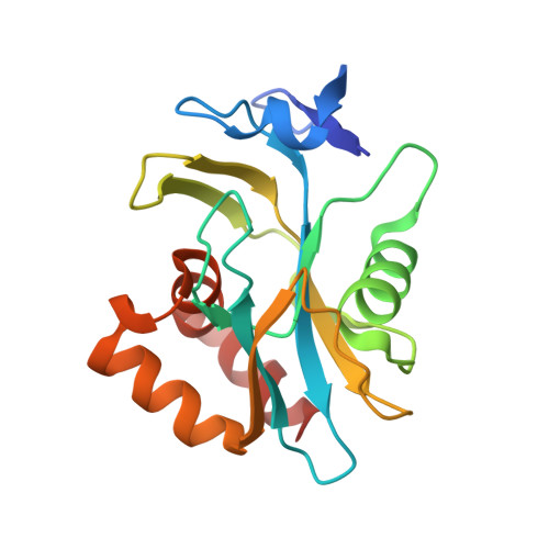
2VNP

Monoclinic form of IDI-1


Experimental Data Snapshot

  • Method: X-RAY DIFFRACTION
  • Resolution: 2.19 Å
  • R-Value Free: 0.255 
  • R-Value Work: 0.194 
  • R-Value Observed: 0.197 

Starting Model: experimental
View more details

wwPDB Validation   3D Report Full Report


Ligand Structure Quality Assessment 


This is version 1.3 of the entry. See complete history


Literature

Monoclinic Form of Isopentenyl Diphosphate Isomerase: A Case of Polymorphism in Biomolecular Crystals.

De Ruyck, J.Oudjama, Y.Wouters, J.

(2008) Acta Crystallogr Sect F Struct Biol Cryst Commun 64: 239

  • DOI: https://doi.org/10.1107/S174430910800568X
  • Primary Citation of Related Structures:  
    2VNP, 2VNQ

  • PubMed Abstract: 

    Type 1 isopentenyl diphosphate isomerase (IDI-1) has been crystallized in a new crystal form. After data collection from small thin needle-shaped crystals, a new monoclinic form of the studied protein was identified. In this article, the three crystal forms of IDI-1 (orthorhombic, monoclinic and trigonal) are compared.


  • Organizational Affiliation

    Laboratoire de Chimie Biologique Structurale, FUNDP University of Namur, 61 Rue de Bruxelles, 5000 Namur, Belgium. jerome.deruyck@fundp.ac.be


Macromolecules
Find similar proteins by:  (by identity cutoff)  |  3D Structure
Entity ID: 1
MoleculeChains Sequence LengthOrganismDetailsImage
ISOPENTENYL-DIPHOSPHATE DELTA-ISOMERASE
A, B
183Escherichia coli BL21Mutation(s): 0 
EC: 5.3.3.2
UniProt
Find proteins for Q46822 (Escherichia coli (strain K12))
Explore Q46822 
Go to UniProtKB:  Q46822
Entity Groups  
Sequence Clusters30% Identity50% Identity70% Identity90% Identity95% Identity100% Identity
UniProt GroupQ46822
Sequence Annotations
Expand
  • Reference Sequence
Small Molecules
Binding Affinity Annotations 
IDSourceBinding Affinity
DED PDBBind:  2VNP Ki: 0.12 (nM) from 1 assay(s)
Experimental Data & Validation

Experimental Data

  • Method: X-RAY DIFFRACTION
  • Resolution: 2.19 Å
  • R-Value Free: 0.255 
  • R-Value Work: 0.194 
  • R-Value Observed: 0.197 
  • Space Group: P 1 21 1
Unit Cell:
Length ( Å )Angle ( ˚ )
a = 43.2α = 90
b = 69.1β = 100.3
c = 62.4γ = 90
Software Package:
Software NamePurpose
REFMACrefinement
XDSdata reduction
XDSdata scaling
PHASERphasing

Structure Validation

View Full Validation Report


Ligand Structure Quality Assessment 


Entry History 

Deposition Data

Revision History  (Full details and data files)

  • Version 1.0: 2008-03-18
    Type: Initial release
  • Version 1.1: 2011-05-08
    Changes: Version format compliance
  • Version 1.2: 2011-07-13
    Changes: Version format compliance
  • Version 1.3: 2023-12-13
    Changes: Data collection, Database references, Derived calculations, Other, Refinement description