2XLN

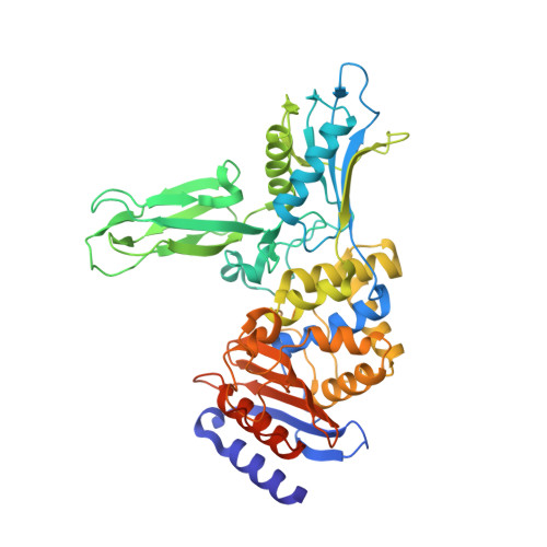
Crystal structure of a complex between Actinomadura R39 DD-peptidase and a boronate inhibitor


Experimental Data Snapshot

  • Method: X-RAY DIFFRACTION
  • Resolution: 2.40 Å
  • R-Value Free: 0.247 
  • R-Value Work: 0.191 
  • R-Value Observed: 0.194 

wwPDB Validation   3D Report Full Report


Ligand Structure Quality Assessment 


This is version 1.3 of the entry. See complete history


Literature

Structure Guided Development of Potent Reversibly Binding Penicillin Binding Protein Inhibitors

Woon, E.C.Y.Zervosen, A.Sauvage, E.Simmons, K.J.Ivec, M.Inglis, S.R.Fishwick, C.W.G.Gobec, S.Charlier, P.Luxen, A.Schofield, C.J.

(2011) ACS Med Chem Lett 2: 219

  • DOI: https://doi.org/10.1021/ml100260x
  • Primary Citation of Related Structures:  
    2XK1, 2XLN

  • PubMed Abstract: 

    Following from the evaluation of different types of electrophiles, combined modeling and crystallographic analyses are used to generate potent boronic acid based inhibitors of a penicillin binding protein. The results suggest that a structurally informed approach to penicillin binding protein inhibition will be useful for the development of both improved reversibly binding inhibitors, including boronic acids, and acylating inhibitors, such as β-lactams.


  • Organizational Affiliation

    Chemistry Research Laboratory, Department of Chemistry, University of Oxford , 12 Mansfield Road, Oxford OX1 3TA, United Kingdom.


Macromolecules
Find similar proteins by:  (by identity cutoff)  |  3D Structure
Entity ID: 1
MoleculeChains Sequence LengthOrganismDetailsImage
D-ALANYL-D-ALANINE CARBOXYPEPTIDASE,
A, B, C, D
489Actinomadura sp. R39Mutation(s): 0 
EC: 3.4.16.4
UniProt
Find proteins for P39045 (Actinomadura sp. (strain R39))
Explore P39045 
Go to UniProtKB:  P39045
Entity Groups  
Sequence Clusters30% Identity50% Identity70% Identity90% Identity95% Identity100% Identity
UniProt GroupP39045
Sequence Annotations
Expand
  • Reference Sequence
Small Molecules
Ligands 3 Unique
IDChains Name / Formula / InChI Key2D Diagram3D Interactions
EWA
Query on EWA

Download Ideal Coordinates CCD File 
E [auth A],
K [auth B],
Q [auth C],
V [auth D]
[(1S)-1-{[(2,6-DIMETHOXYPHENYL)CARBONYL]AMINO}ETHYL]BORONIC ACID
C11 H16 B N O5
DXKXQRZEPYKFAT-SSDOTTSWSA-N
SO4
Query on SO4

Download Ideal Coordinates CCD File 
F [auth A]
G [auth A]
H [auth A]
I [auth A]
L [auth B]
F [auth A],
G [auth A],
H [auth A],
I [auth A],
L [auth B],
M [auth B],
N [auth B],
O [auth B],
P [auth B],
R [auth C],
S [auth C],
T [auth C],
U [auth C],
W [auth D],
X [auth D],
Y [auth D],
Z [auth D]
SULFATE ION
O4 S
QAOWNCQODCNURD-UHFFFAOYSA-L
CO
Query on CO

Download Ideal Coordinates CCD File 
AA [auth D],
J [auth A]
COBALT (II) ION
Co
XLJKHNWPARRRJB-UHFFFAOYSA-N
Binding Affinity Annotations 
IDSourceBinding Affinity
EWA PDBBind:  2XLN IC50: 3.30e+4 (nM) from 1 assay(s)
Experimental Data & Validation

Experimental Data

  • Method: X-RAY DIFFRACTION
  • Resolution: 2.40 Å
  • R-Value Free: 0.247 
  • R-Value Work: 0.191 
  • R-Value Observed: 0.194 
  • Space Group: P 1 21 1
Unit Cell:
Length ( Å )Angle ( ˚ )
a = 102.994α = 90
b = 91.258β = 94.46
c = 106.878γ = 90
Software Package:
Software NamePurpose
REFMACrefinement
MOSFLMdata reduction
SCALAdata scaling

Structure Validation

View Full Validation Report


Ligand Structure Quality Assessment 


Entry History 

Deposition Data

Revision History  (Full details and data files)

  • Version 1.0: 2011-01-26
    Type: Initial release
  • Version 1.1: 2012-06-06
    Changes: Database references, Version format compliance
  • Version 1.2: 2014-06-18
    Changes: Database references
  • Version 1.3: 2024-11-06
    Changes: Data collection, Database references, Derived calculations, Other, Structure summary