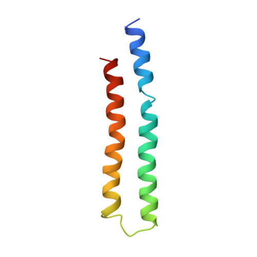
2XOK

Refined structure of yeast F1c10 ATPase complex to 3 A resolution


Experimental Data Snapshot

  • Method: X-RAY DIFFRACTION
  • Resolution: 3.01 Å
  • R-Value Free: 0.253 
  • R-Value Work: 0.210 
  • R-Value Observed: 0.211 

Starting Model: experimental
View more details

wwPDB Validation   3D Report Full Report


Ligand Structure Quality Assessment 


This is version 1.5 of the entry. See complete history


Literature

Molecular architecture of the rotary motor in ATP synthase.

Stock, D.Leslie, A.G.Walker, J.E.

(1999) Science 286: 1700-1705

  • DOI: https://doi.org/10.1126/science.286.5445.1700
  • Primary Citation of Related Structures:  
    1QO1, 2XOK

  • PubMed Abstract: 

    Adenosine triphosphate (ATP) synthase contains a rotary motor involved in biological energy conversion. Its membrane-embedded F0 sector has a rotation generator fueled by the proton-motive force, which provides the energy required for the synthesis of ATP by the F1 domain. An electron density map obtained from crystals of a subcomplex of yeast mitochondrial ATP synthase shows a ring of 10 c subunits. Each c subunit forms an alpha-helical hairpin. The interhelical loops of six to seven of the c subunits are in close contact with the gamma and delta subunits of the central stalk. The extensive contact between the c ring and the stalk suggests that they may rotate as an ensemble during catalysis.


  • Organizational Affiliation

    Medical Research Council Dunn Human Nutrition Unit, Hills Road, Cambridge CB2 2XY, UK.


Macromolecules
Find similar proteins by:  (by identity cutoff)  |  3D Structure
Entity ID: 1
MoleculeChains Sequence LengthOrganismDetailsImage
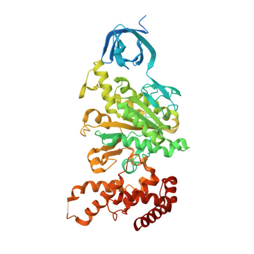
ATP SYNTHASE SUBUNIT ALPHA, MITOCHONDRIAL
A, B, C
545Saccharomyces cerevisiaeMutation(s): 0 
UniProt
Find proteins for P07251 (Saccharomyces cerevisiae (strain ATCC 204508 / S288c))
Go to UniProtKB:  P07251
Sequence Annotations
Expand
  • Reference Sequence
Find similar proteins by:  (by identity cutoff)  |  3D Structure
Entity ID: 2
MoleculeChains Sequence LengthOrganismDetailsImage
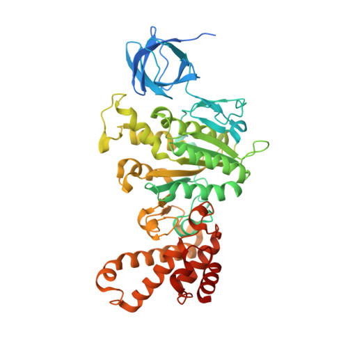
ATP SYNTHASE SUBUNIT BETA, MITOCHONDRIAL
D, E, F
511Saccharomyces cerevisiaeMutation(s): 0 
UniProt
Find proteins for P00830 (Saccharomyces cerevisiae (strain ATCC 204508 / S288c))
Go to UniProtKB:  P00830
Sequence Annotations
Expand
  • Reference Sequence
Find similar proteins by:  (by identity cutoff)  |  3D Structure
Entity ID: 3
MoleculeChains Sequence LengthOrganismDetailsImage
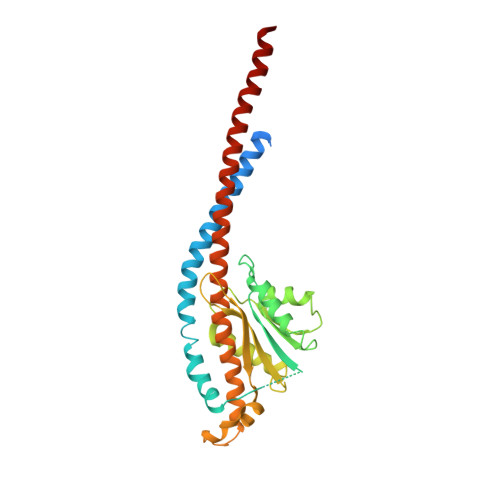
ATP SYNTHASE SUBUNIT GAMMA, MITOCHONDRIAL311Saccharomyces cerevisiaeMutation(s): 0 
UniProt
Find proteins for P38077 (Saccharomyces cerevisiae (strain ATCC 204508 / S288c))
Go to UniProtKB:  P38077
Sequence Annotations
Expand
  • Reference Sequence
Find similar proteins by:  (by identity cutoff)  |  3D Structure
Entity ID: 4
MoleculeChains Sequence LengthOrganismDetailsImage
ATP SYNTHASE160Saccharomyces cerevisiaeMutation(s): 0 
UniProt
Find proteins for Q12165 (Saccharomyces cerevisiae (strain ATCC 204508 / S288c))
Go to UniProtKB:  Q12165
Sequence Annotations
Expand
  • Reference Sequence
Find similar proteins by:  (by identity cutoff)  |  3D Structure
Entity ID: 5
MoleculeChains Sequence LengthOrganismDetailsImage
ATP SYNTHASE CATALYTIC SECTOR F1 EPSILON SUBUNIT61Saccharomyces cerevisiaeMutation(s): 0 
UniProt
Find proteins for E9P9X4 (Saccharomyces cerevisiae)
Go to UniProtKB:  E9P9X4
Sequence Annotations
Expand
  • Reference Sequence
Find similar proteins by:  (by identity cutoff)  |  3D Structure
Entity ID: 6
MoleculeChains Sequence LengthOrganismDetailsImage
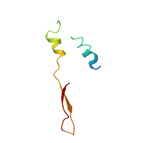
ATP SYNTHASE SUBUNIT 9, MITOCHONDRIAL76Saccharomyces cerevisiaeMutation(s): 0 
UniProt
Find proteins for P61829 (Saccharomyces cerevisiae (strain ATCC 204508 / S288c))
Go to UniProtKB:  P61829
Sequence Annotations
Expand
  • Reference Sequence
Small Molecules
Ligands 2 Unique
IDChains Name / Formula / InChI Key2D Diagram3D Interactions
ANP
Query on ANP

Download Ideal Coordinates CCD File 
BA [auth F],
T [auth A],
V [auth B],
X [auth C],
Z [auth D]
PHOSPHOAMINOPHOSPHONIC ACID-ADENYLATE ESTER
C10 H17 N6 O12 P3
PVKSNHVPLWYQGJ-KQYNXXCUSA-N
MG
Query on MG

Download Ideal Coordinates CCD File 
AA [auth D],
CA [auth F],
U [auth A],
W [auth B],
Y [auth C]
MAGNESIUM ION
Mg
JLVVSXFLKOJNIY-UHFFFAOYSA-N
Experimental Data & Validation

Experimental Data

  • Method: X-RAY DIFFRACTION
  • Resolution: 3.01 Å
  • R-Value Free: 0.253 
  • R-Value Work: 0.210 
  • R-Value Observed: 0.211 
  • Space Group: P 1 21 1
Unit Cell:
Length ( Å )Angle ( ˚ )
a = 135.352α = 90
b = 173.711β = 91.77
c = 137.889γ = 90
Software Package:
Software NamePurpose
REFMACrefinement
MOSFLMdata reduction
SCALAdata scaling
AMoREphasing

Structure Validation

View Full Validation Report


Ligand Structure Quality Assessment 


Entry History 

Deposition Data

Revision History  (Full details and data files)

  • Version 1.0: 2010-09-22
    Type: Initial release
  • Version 1.1: 2011-05-08
    Changes: Version format compliance
  • Version 1.2: 2011-07-13
    Changes: Version format compliance
  • Version 1.3: 2019-05-08
    Changes: Data collection, Experimental preparation, Other
  • Version 1.4: 2019-10-09
    Changes: Data collection, Database references, Other
  • Version 1.5: 2023-12-20
    Changes: Data collection, Database references, Derived calculations, Refinement description