2Z7L | pdb_00002z7l

Unphosphorylated Mitogen Activated Protein Kinase ERK2 in Complex with (4-{[5-Carbamoyl-4-(3-Methylanilino)Pyrimidin 2-Yl]Amino}Phenyl)Acetic Acid


Experimental Data Snapshot

  • Method: X-RAY DIFFRACTION
  • Resolution: 2.41 Å
  • R-Value Free: 
    0.276 (Depositor), 0.269 (DCC) 
  • R-Value Work: 
    0.223 (Depositor), 0.219 (DCC) 

Starting Model: experimental
View more details

wwPDB Validation 3D Report Full Report

Validation slider image for 2Z7L

Ligand Structure Quality Assessment 


This is version 1.2 of the entry. See complete history

Literature

Identification of a key element for hydrogen-bonding patterns between protein kinases and their inhibitors

Katayama, N.Orita, M.Yamaguchi, T.Hisamichi, H.Kuromitsu, S.Kurihara, H.Sakashita, H.Matsumoto, Y.Fujita, S.Niimi, T.

(2008) Proteins 73: 795-801

  • DOI: https://doi.org/10.1002/prot.22207
  • Primary Citation Related Structures: 
    2Z7L, 2Z8C

  • PubMed Abstract: 

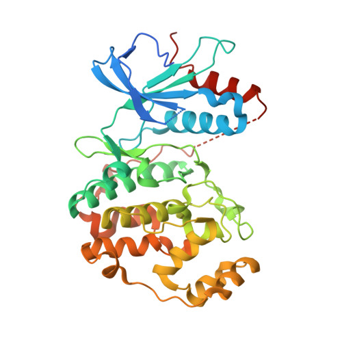
    In this article, we report crystal structures for inhibitor-kinase complexes in which the inhibitor has different binding orientations and hydrogen-bonding patterns with extracellular-signal regulated kinase 2 and insulin receptor tyrosine kinase. Our crystallographic studies, and sequence and structural analyses of 532 coordinates of kinases held in the Protein Data Bank, suggest that the length of the "specificity linker" described here is a key structural element of the hydrogen-bonding patterns between protein kinases and their inhibitors.


  • Organizational Affiliation
    • Drug Discovery Research, Astellas Pharma Inc., 21 Miyukigaoka, Tsukuba, Ibaraki 305-8585, Japan.

Macromolecule Content 

  • Total Structure Weight: 43.07 kDa 
  • Atom Count: 2,864 
  • Modeled Residue Count: 329 
  • Deposited Residue Count: 366 
  • Unique protein chains: 1

Macromolecules

Find similar proteins by:|  3D Structure
Entity ID: 1
MoleculeChains  Sequence LengthOrganismDetailsImage
Mitogen-activated protein kinase 1366Rattus norvegicusMutation(s): 0 
Gene Names: Mapk1Erk2MapkPrkm1
EC: 2.7.11.24
UniProt
Find proteins for P63086 (Rattus norvegicus)
Explore P63086 
Go to UniProtKB:  P63086
Entity Groups
Sequence Clusters30% Identity50% Identity70% Identity90% Identity95% Identity100% Identity
UniProt GroupP63086
Sequence Annotations
Expand
Reference Sequence

Experimental Data & Validation

Experimental Data

  • Method: X-RAY DIFFRACTION
  • Resolution: 2.41 Å
  • R-Value Free:  0.276 (Depositor), 0.269 (DCC) 
  • R-Value Work:  0.223 (Depositor), 0.219 (DCC) 
Space Group: P 1 21 1
Unit Cell:
Length ( Å )Angle ( ˚ )
a = 48.873α = 90
b = 69.962β = 109.58
c = 60.775γ = 90
Software Package:
Software NamePurpose
CNXrefinement
MOSFLMdata reduction
SCALAdata scaling
CNXphasing

Structure Validation

View Full Validation Report


Ligand Structure Quality Assessment 


Entry History 

Deposition Data

Revision History  (Full details and data files)

  • Version 1.0: 2008-08-12
    Type: Initial release
  • Version 1.1: 2011-07-13
    Changes: Version format compliance
  • Version 1.2: 2023-11-01
    Changes: Data collection, Database references, Derived calculations, Refinement description