2ZI0

Crystal structure of Tav2b/siRNA complex


Experimental Data Snapshot

  • Method: X-RAY DIFFRACTION
  • Resolution: 2.82 Å
  • R-Value Free: 0.276 
  • R-Value Work: 0.214 
  • R-Value Observed: 0.216 

wwPDB Validation   3D Report Full Report


This is version 1.2 of the entry. See complete history


Literature

Structural basis for RNA-silencing suppression by Tomato aspermy virus protein 2b

Chen, H.-Y.Yang, J.Lin, C.Yuan, Y.A.

(2008) EMBO Rep 9: 754-760

  • DOI: https://doi.org/10.1038/embor.2008.118
  • Primary Citation of Related Structures:  
    2ZI0

  • PubMed Abstract: 

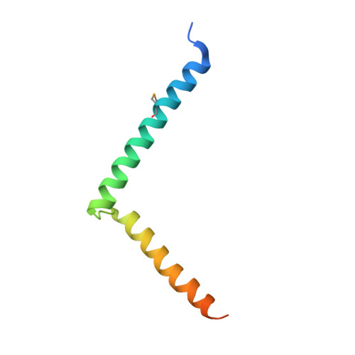
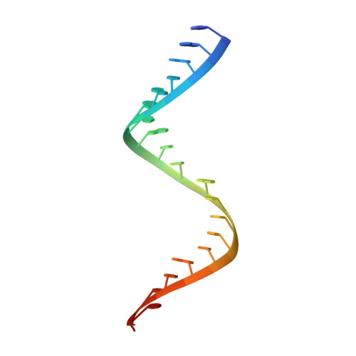
    The 2b proteins encoded by cucumovirus act as post-transcriptional gene silencing suppressors to counter host defence during infection. Here we report the crystal structure of Tomato aspermy virus 2b (TAV2b) protein bound to a 19 bp small interfering RNA (siRNA) duplex. TAV2b adopts an all alpha-helix structure and forms a homodimer to measure siRNA duplex in a length-preference mode. TAV2b has a pair of hook-like structures to recognize simultaneously two alpha-helical turns of A-form RNA duplex by fitting its alpha-helix backbone into two adjacent major grooves of siRNA duplex. The conserved pi-stackings between tryptophan and the 5'-terminal base of siRNA duplex from both ends enhance the recognition. TAV2b further oligomerizes to form a dimer of dimers through the conserved leucine-zipper-like motif at its amino-terminal alpha-helix. Biochemical experiments suggest that TAV2b might interfere with the post-transcriptional gene silencing pathway by directly binding to siRNA duplex.


  • Organizational Affiliation

    Host-Pathogen Interaction Group, National University of Singapore, Singapore.


Macromolecules

Find similar proteins by:  (by identity cutoff)  |  3D Structure
Entity ID: 1
MoleculeChains Sequence LengthOrganismDetailsImage
Protein 2b
A, B
75Tomato aspermy virusMutation(s): 0 
Gene Names: RNA4A
UniProt
Find proteins for Q8UYT3 (Tomato aspermy virus)
Explore Q8UYT3 
Go to UniProtKB:  Q8UYT3
Entity Groups  
Sequence Clusters30% Identity50% Identity70% Identity90% Identity95% Identity100% Identity
UniProt GroupQ8UYT3
Sequence Annotations
Expand
  • Reference Sequence

Find similar nucleic acids by:  Sequence   |   3D Structure  

Entity ID: 2
MoleculeChains LengthOrganismImage
RNA (5'-D(P*AP*GP*AP*CP*AP*GP*CP*AP*UP*UP*AP*UP*GP*CP*UP*GP*UP*CP*UP*UP*U)-3')
C, D
21N/A
Sequence Annotations
Expand
  • Reference Sequence
Small Molecules
Modified Residues  1 Unique
IDChains TypeFormula2D DiagramParent
MSE
Query on MSE
A, B
L-PEPTIDE LINKINGC5 H11 N O2 SeMET
Experimental Data & Validation

Experimental Data

  • Method: X-RAY DIFFRACTION
  • Resolution: 2.82 Å
  • R-Value Free: 0.276 
  • R-Value Work: 0.214 
  • R-Value Observed: 0.216 
  • Space Group: P 21 21 2
Unit Cell:
Length ( Å )Angle ( ˚ )
a = 86.455α = 90
b = 122.043β = 90
c = 28.193γ = 90
Software Package:
Software NamePurpose
REFMACrefinement
HKL-2000data collection
HKL-2000data reduction
HKL-2000data scaling
SHARPphasing

Structure Validation

View Full Validation Report


Entry History 

Deposition Data

Revision History  (Full details and data files)

  • Version 1.0: 2008-07-22
    Type: Initial release
  • Version 1.1: 2011-07-13
    Changes: Advisory, Version format compliance
  • Version 1.2: 2024-10-16
    Changes: Data collection, Database references, Derived calculations, Structure summary