Interaction between left-handed Z-DNA and polyamine - 3. The crystal structure of the d(CG)3 and thermospermine complex.
Ohishi, H., Terasoma, N., Nakanishi, I., van der Marel, G., van Boom, J.H., Rich, A., Wang, A.H., Hakoshima, T., Tomita, K.(1996) FEBS Lett 398: 291-296
- PubMed: 8977125 Search on PubMed
- DOI: https://doi.org/10.1016/s0014-5793(96)01225-2
- Primary Citation Related Structures:
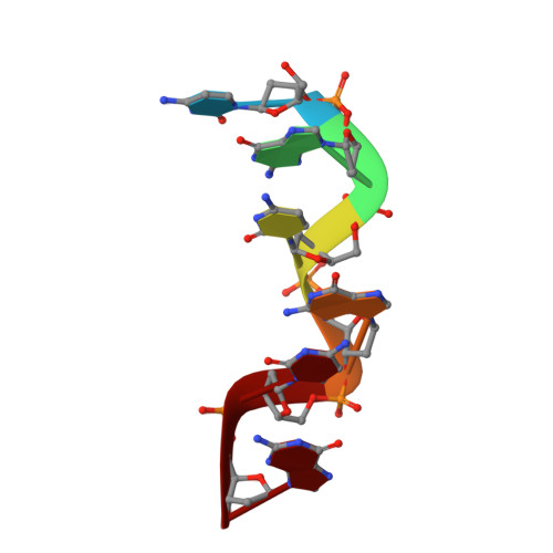
336D - PubMed Abstract:
The DNA fragment, d(CG)3, was co-crystallized with N-(3-amino-propyl)-N-(5-aminopropyl)-l,4 -diaminobutane (thermospermine; PA(334)), a polyamine metabolized from the nucleic acid. By using a good crystal with dimensions of 0.5 x 0.5 x 0.5 mm3, X-ray intensity data were collected up to 1.0 A resolution. Two thermospermine molecules and a magnesium cation were bound to the left-handed double-helical d(CG)3 molecule. The d(CG)3 molecule adopted the left-handed Z-conformation and two thermospermine molecules and a magnesium cation neutralized the negative charges of the phosphate groups of the d(CG)3 molecule. Furthermore, the binding modes between d(CG)3 and thermospermine were different from those of d(CG)3 complexes with PA(24), spermidine and spermine. This is the first case in which it was determined by X-ray crystallographic analysis that one of two thermospermine molecules bound three d(CG)3 duplexes which were symmetrically related to each other, and the other formed two hydrogen bonds at the N(5) and N(9) atoms with two adjacent nucleotide phosphate groups of a single d(CG)3 strand at the minor groove. Furthermore, no direct coordination bond was found between the d(CG)3 molecule and the magnesium cation.
- Osaka University of Pharmaceutical Sciences, Takatsuki, Japan. ohishi@oysun01.oups.ac.jp
Organizational Affiliation: