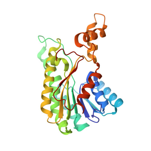
3AI3

The crystal structure of L-Sorbose reductase from Gluconobacter frateurii complexed with NADPH and L-sorbose


Experimental Data Snapshot

  • Method: X-RAY DIFFRACTION
  • Resolution: 1.80 Å
  • R-Value Free: 0.249 
  • R-Value Work: 0.212 
  • R-Value Observed: 0.214 

Starting Model: experimental
View more details

wwPDB Validation   3D Report Full Report


Ligand Structure Quality Assessment 


This is version 1.4 of the entry. See complete history


Literature

The Crystal Structure of l-Sorbose Reductase from Gluconobacter frateurii Complexed with NADPH and l-Sorbose

Kubota, K.Nagata, K.Okai, M.Miyazono, K.Soemphol, W.Ohtsuka, J.Yamamura, A.Saichana, N.Toyama, H.Matsushita, K.Tanokura, M.

(2011) J Mol Biol 407: 543-555

  • DOI: https://doi.org/10.1016/j.jmb.2011.01.008
  • Primary Citation of Related Structures:  
    3AI1, 3AI2, 3AI3

  • PubMed Abstract: 

    l-Sorbose reductase from Gluconobacter frateurii (SR) is an NADPH-dependent oxidoreductase. SR preferentially catalyzes the reversible reaction between d-sorbitol and l-sorbose with high substrate specificity. To elucidate the structural basis of the catalytic mechanism and the substrate specificity of SR, we have determined the structures of apo-SR, SR in complex with NADPH, and the inactive mutant (His116Leu) of SR in complex with NADPH and l-sorbose at 2.83 Å, 1.90 Å, and 1.80 Å resolutions, respectively. Our results show that SR belongs to the short-chain dehydrogenase/reductase (SDR) family and forms a tetrameric structure. Although His116 is not conserved among SDR family enzymes, the structures of SR have revealed that His116 is important for the stabilization of the proton relay system and for active-site conformation as a fourth catalytic residue. In the ternary complex structure, l-sorbose is recognized by 11 hydrogen bonds. Site-directed mutagenesis of residues around the l-sorbose-binding site has shown that the loss of almost full enzymatic activity was caused by not only the substitution of putative catalytic residues but also the substitution of the residue used for the recognition of the C4 hydroxyl groups of l-sorbose (Glu154) and of the residues used for the construction of the substrate-binding pocket (Cys146 and Gly188). The recognition of the C4 hydroxyl group of l-sorbose would be indispensable for the substrate specificity of SR, which recognizes only l-sorbose and d-sorbitol but not other sugars. Our results indicated that these residues were crucial for the substrate recognition and specificity of SR.


  • Organizational Affiliation

    Department of Applied Biological Chemistry, Graduate School of Agricultural and Life Sciences, University of Tokyo, 1-1-1 Yayoi, Bunkyo-ku, Tokyo 113-8657, Japan.


Macromolecules
Find similar proteins by:  (by identity cutoff)  |  3D Structure
Entity ID: 1
MoleculeChains Sequence LengthOrganismDetailsImage
NADPH-sorbose reductaseA,
B [auth C],
C [auth E],
D [auth G]
263Gluconobacter frateuriiMutation(s): 1 
Gene Names: sboA
EC: 1.1.1.289
UniProt
Find proteins for A4PB64 (Gluconobacter frateurii)
Explore A4PB64 
Go to UniProtKB:  A4PB64
Entity Groups  
Sequence Clusters30% Identity50% Identity70% Identity90% Identity95% Identity100% Identity
UniProt GroupA4PB64
Sequence Annotations
Expand
  • Reference Sequence
Small Molecules
Ligands 3 Unique
IDChains Name / Formula / InChI Key2D Diagram3D Interactions
NDP
Query on NDP

Download Ideal Coordinates CCD File 
E [auth A],
G [auth C],
I [auth E],
M [auth G]
NADPH DIHYDRO-NICOTINAMIDE-ADENINE-DINUCLEOTIDE PHOSPHATE
C21 H30 N7 O17 P3
ACFIXJIJDZMPPO-NNYOXOHSSA-N
SOE
Query on SOE

Download Ideal Coordinates CCD File 
K [auth E],
O [auth G],
P [auth G],
Q [auth G]
alpha-L-sorbopyranose
C6 H12 O6
LKDRXBCSQODPBY-BGPJRJDNSA-N
SOL
Query on SOL

Download Ideal Coordinates CCD File 
F [auth A],
H [auth C],
J [auth E],
L [auth E],
N [auth G]
L-sorbose
C6 H12 O6
BJHIKXHVCXFQLS-OTWZMJIISA-N
Experimental Data & Validation

Experimental Data

  • Method: X-RAY DIFFRACTION
  • Resolution: 1.80 Å
  • R-Value Free: 0.249 
  • R-Value Work: 0.212 
  • R-Value Observed: 0.214 
  • Space Group: P 21 21 21
Unit Cell:
Length ( Å )Angle ( ˚ )
a = 61.04α = 90
b = 124.41β = 90
c = 124.54γ = 90
Software Package:
Software NamePurpose
ADSCdata collection
REFMACrefinement
XDSdata reduction
XDSdata scaling

Structure Validation

View Full Validation Report


Ligand Structure Quality Assessment 


Entry History 

Deposition Data

Revision History  (Full details and data files)

  • Version 1.0: 2011-02-09
    Type: Initial release
  • Version 1.1: 2011-07-13
    Changes: Version format compliance
  • Version 1.2: 2013-11-20
    Changes: Non-polymer description
  • Version 1.3: 2020-07-29
    Type: Remediation
    Reason: Carbohydrate remediation
    Changes: Data collection, Database references, Derived calculations
  • Version 1.4: 2023-11-01
    Changes: Data collection, Database references, Refinement description, Structure summary