FYX-051: A Novel and Potent Hybrid-Type Inhibitor of Xanthine Oxidoreductase
Matsumoto, K., Okamoto, K., Ashizawa, N., Nishino, T.(2011) J Pharmacol Exp Ther 336: 95-103
- PubMed: 20952484 Search on PubMed
- DOI: https://doi.org/10.1124/jpet.110.174540
- Primary Citation Related Structures:
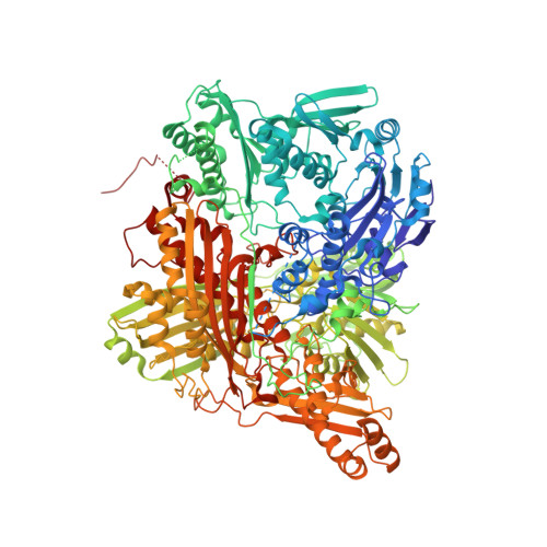
3AM9 - PubMed Abstract:
4-[5-(Pyridin-4-yl)-1H-1,2,4-triazol-3-yl]pyridine-2-carbonitrile (FYX-051) is a potent inhibitor of bovine milk xanthine oxidoreductase (XOR). Steady-state kinetics study showed that it initially behaved as a competitive-type inhibitor with a K(i) value of 5.7 × 10(-9) M, then after a few minutes it formed a tight complex with XOR via a Mo-oxygen-carbon atom covalent linkage, as reported previously (Proc Natl Acad Sci USA 101:7931-7936, 2004). Thus, FYX-051 is a hybrid-type inhibitor exhibiting both structure- and mechanism-based inhibition. The FYX-051-XOR complex decomposed with a half-life of 20.4 h, but the enzyme activity did not fully recover. This was found to be caused by XOR-mediated conversion of FYX-051 to 4-[5-(2-hydroxypyridin-4-yl)-1H-1,2,4-triazol-3-yl]pyridine-2-carbonitrile (2-hydroxy-FYX-051), as well as formation of 6-hydroxy-4-[5-(2-hydroxypyridin-4-yl)-1H-1,2,4-triazol-3-yl]pyridine-2-carbonitrile (dihydroxy-FYX-051) and 4-[5-(2,6-dihydroxypyridin-4-yl)-1H-1,2,4-triazol-3-yl]-6-hydroxypyridine-2-carbonitrile (trihydroxy-FYX-051) during prolonged incubation for up to 72 h. A distinct charge-transfer band was observed concomitantly with the formation of the trihydroxy-FYX-051-XOR complex. Crystallographic analysis of the charge-transfer complex indicated that a Mo-nitrogen-carbon bond was formed between molybdenum of XOR and the nitrile group of trihydroxy-FYX-051. FYX-051 showed a potent and long-lasting hypouricemic effect in a rat model of potassium oxonate-induced hyperuricemia, and it seems to be a promising candidate for the clinical treatment of hyperuricemia.
- Department of Biochemistry and Molecular Biology, Nippon Medical School, 1-1-5 Sendagi, Bunkyo-ku, Tokyo 113-8602, Japan.
Organizational Affiliation: