3AVQ | pdb_00003avq

Pantothenate kinase from Mycobacterium tuberculosis (MtPanK) in complex with N9-Pan


Experimental Data Snapshot

  • Method: X-RAY DIFFRACTION
  • Resolution: 3.00 Å
  • R-Value Free: 
    0.260 (Depositor), 0.260 (DCC) 
  • R-Value Work: 
    0.204 (Depositor), 0.204 (DCC) 
  • R-Value Observed: 
    0.204 (Depositor) 

Starting Model: experimental
View more details

wwPDB Validation 3D Report Full Report

Validation slider image for 3AVQ

Ligand Structure Quality Assessment 


This is version 1.2 of the entry. See complete history

Literature

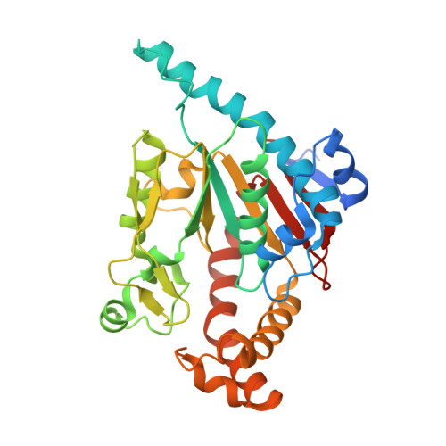
Location and conformation of pantothenate and its derivatives in Mycobacterium tuberculosis pantothenate kinase: insights into enzyme action

Chetnani, B.Kumar, P.Abhinav, K.V.Chhibber, M.Surolia, A.Vijayan, M.

(2011) Acta Crystallogr D Biol Crystallogr 67: 774-783

  • DOI: https://doi.org/10.1107/S0907444911024462
  • Primary Citation Related Structures: 
    3AVO, 3AVP, 3AVQ

  • PubMed Abstract: 

    Previous studies of complexes of Mycobacterium tuberculosis PanK (MtPanK) with nucleotide diphosphates and nonhydrolysable analogues of nucleoside triphosphates in the presence or the absence of pantothenate established that the enzyme has dual specificity for ATP and GTP, revealed the unusual movement of ligands during enzyme action and provided information on the effect of pantothenate on the location and conformation of the nucleotides at the beginning and the end of enzyme action. The X-ray analyses of the binary complexes of MtPanK with pantothenate, pantothenol and N-nonylpantothenamide reported here demonstrate that in the absence of nucleotide these ligands occupy, with a somewhat open conformation, a location similar to that occupied by phosphopantothenate in the `end' complexes, which differs distinctly from the location of pantothenate in the closed conformation in the ternary `initiation' complexes. The conformation and the location of the nucleotide were also different in the initiation and end complexes. An invariant arginine appears to play a critical role in the movement of ligands that takes place during enzyme action. The work presented here completes the description of the locations and conformations of nucleoside diphosphates and triphosphates and pantothenate in different binary and ternary complexes, and suggests a structural rationale for the movement of ligands during enzyme action. The present investigation also suggests that N-alkylpantothenamides could be phosphorylated by the enzyme in the same manner as pantothenate.


  • Organizational Affiliation
    • Molecular Biophysics Unit, Indian Institute of Science, Bangalore.

Macromolecule Content 

  • Total Structure Weight: 38.26 kDa 
  • Atom Count: 2,753 
  • Modeled Residue Count: 307 
  • Deposited Residue Count: 322 
  • Unique protein chains: 1

Macromolecules

Find similar proteins by:|  3D Structure
Entity ID: 1
MoleculeChains  Sequence LengthOrganismDetailsImage
Pantothenate kinase322Mycobacterium tuberculosisMutation(s): 0 
Gene Names: coaARv1092c
EC: 2.7.1.33
UniProt
Find proteins for P9WPA7 (Mycobacterium tuberculosis (strain ATCC 25618 / H37Rv))
Explore P9WPA7 
Go to UniProtKB:  P9WPA7
Entity Groups
Sequence Clusters30% Identity50% Identity70% Identity90% Identity95% Identity100% Identity
UniProt GroupP9WPA7
Sequence Annotations
Expand
Reference Sequence

Small Molecules

Ligands 3 Unique
IDChains Name / Formula / InChI Key2D Diagram3D Interactions
MV1

Query on MV1


Download:Ideal Coordinates CCD File
B [auth A](2S)-2,4-dihydroxy-3,3-dimethyl-N-[3-(nonylamino)-3-oxopropyl]butanamide
C18 H36 N2 O4
OAZREYBYPROKTB-MRXNPFEDSA-N
FLC

Query on FLC


Download:Ideal Coordinates CCD File
C [auth A]CITRATE ANION
C6 H5 O7
KRKNYBCHXYNGOX-UHFFFAOYSA-K
GOL

Query on GOL


Download:Ideal Coordinates CCD File
D [auth A]
E [auth A]
F [auth A]
G [auth A]
H [auth A]
D [auth A],
E [auth A],
F [auth A],
G [auth A],
H [auth A],
I [auth A],
J [auth A],
K [auth A],
L [auth A]
GLYCEROL
C3 H8 O3
PEDCQBHIVMGVHV-UHFFFAOYSA-N

Experimental Data & Validation

Experimental Data

  • Method: X-RAY DIFFRACTION
  • Resolution: 3.00 Å
  • R-Value Free:  0.260 (Depositor), 0.260 (DCC) 
  • R-Value Work:  0.204 (Depositor), 0.204 (DCC) 
  • R-Value Observed: 0.204 (Depositor) 
Space Group: P 31 2 1
Unit Cell:
Length ( Å )Angle ( ˚ )
a = 103.848α = 90
b = 103.848β = 90
c = 91.031γ = 120
Software Package:
Software NamePurpose
MAR345dtbdata collection
AMoREphasing
CNSrefinement
DENZOdata reduction
SCALEPACKdata scaling

Structure Validation

View Full Validation Report


Ligand Structure Quality Assessment 


Entry History 

Deposition Data

Revision History  (Full details and data files)

  • Version 1.0: 2011-08-17
    Type: Initial release
  • Version 1.1: 2011-11-02
    Changes: Database references, Structure summary
  • Version 1.2: 2023-11-01
    Changes: Data collection, Database references, Derived calculations, Refinement description