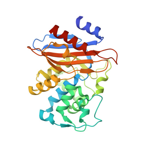
3B3X

Crystal structure of class A beta-lactamase of Bacillus licheniformis BS3 with aminocitrate


Experimental Data Snapshot

  • Method: X-RAY DIFFRACTION
  • Resolution: 2.50 Å
  • R-Value Free: 0.288 
  • R-Value Work: 0.220 
  • R-Value Observed: 0.223 

Starting Model: experimental
View more details

wwPDB Validation   3D Report Full Report


Ligand Structure Quality Assessment 


This is version 1.2 of the entry. See complete history


Literature

2-Aminopropane-1,2,3-tricarboxylic acid: Synthesis and co-crystallization with the class A beta-lactamase BS3 of Bacillus licheniformis

Beck, J.Sauvage, E.Charlier, P.Marchand-Brynaert, J.

(2008) Bioorg Med Chem Lett 18: 3764-3768

  • DOI: https://doi.org/10.1016/j.bmcl.2008.05.045
  • Primary Citation of Related Structures:  
    3B3X

  • PubMed Abstract: 

    The title compound 4 has been prepared in four steps from ethylglycinate in 63% overall yield. This amino analog of citric acid has been co-crystallized with the class A beta-lactamase BS3 of Bacillus licheniformis and the structure of the complex fully analyzed by X-ray diffraction. Tris-ethyl aminocitrate 3 and the free tris-acid 4 have been tested against a member beta-lactamase from all distinct subgroups. They are novel inhibitors of class A beta-lactamases, still modest but more potent than citrate and isocitrate.


  • Organizational Affiliation

    Unité de Chimie Organique et Médicinale, Université catholique de Louvain, Bâtiment Lavoisier, Place Louis Pasteur 1, B-1348 Louvain-la-Neuve, Belgium.


Macromolecules
Find similar proteins by:  (by identity cutoff)  |  3D Structure
Entity ID: 1
MoleculeChains Sequence LengthOrganismDetailsImage
Beta-lactamase
A, B
265Bacillus licheniformisMutation(s): 0 
EC: 3.5.2.6
UniProt
Find proteins for P00808 (Bacillus licheniformis)
Explore P00808 
Go to UniProtKB:  P00808
Entity Groups  
Sequence Clusters30% Identity50% Identity70% Identity90% Identity95% Identity100% Identity
UniProt GroupP00808
Sequence Annotations
Expand
  • Reference Sequence
Small Molecules
Binding Affinity Annotations 
IDSourceBinding Affinity
A33 PDBBind:  3B3X Ki: 2.50e+5 (nM) from 1 assay(s)
Experimental Data & Validation

Experimental Data

  • Method: X-RAY DIFFRACTION
  • Resolution: 2.50 Å
  • R-Value Free: 0.288 
  • R-Value Work: 0.220 
  • R-Value Observed: 0.223 
  • Space Group: P 1 21 1
Unit Cell:
Length ( Å )Angle ( ˚ )
a = 46.665α = 90
b = 104.71β = 93.96
c = 63.884γ = 90
Software Package:
Software NamePurpose
MOSFLMdata reduction
SCALAdata scaling
REFMACrefinement
PDB_EXTRACTdata extraction

Structure Validation

View Full Validation Report


Ligand Structure Quality Assessment 


Entry History 

Deposition Data

Revision History  (Full details and data files)

  • Version 1.0: 2007-11-13
    Type: Initial release
  • Version 1.1: 2011-07-13
    Changes: Version format compliance
  • Version 1.2: 2023-11-01
    Changes: Data collection, Database references, Derived calculations, Refinement description