3E6P | pdb_00003e6p

Crystal structure of human meizothrombin desF1


Experimental Data Snapshot

  • Method: X-RAY DIFFRACTION
  • Resolution: 2.10 Å
  • R-Value Free: 
    0.249 (Depositor), 0.240 (DCC) 
  • R-Value Work: 
    0.211 (Depositor), 0.210 (DCC) 
  • R-Value Observed: 
    0.213 (Depositor) 

Starting Models: experimental
View more details

wwPDB Validation   3D Report Full Report


Ligand Structure Quality Assessment 

Created with Raphaël 2.3.0Worse 01 BetterLigand structure goodness of fit to experimental dataBest fitted DFKClick on this verticalbar to view details

This is version 2.2 of the entry. See complete history


Literature

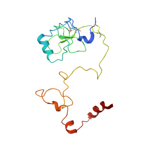
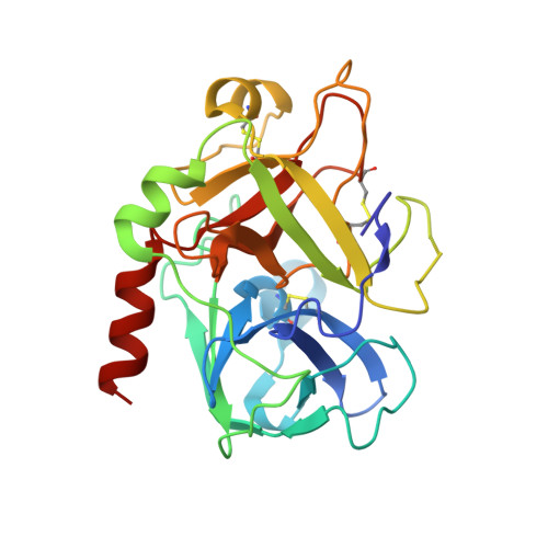
Na(+) binding to meizothrombin desF1.

Papaconstantinou, M.E.Gandhi, P.S.Chen, Z.Bah, A.Di Cera, E.

(2008) Cell Mol Life Sci 65: 3688-3697

  • DOI: https://doi.org/10.1007/s00018-008-8502-7
  • Primary Citation of Related Structures:  
    3E6P

  • PubMed Abstract: 

    Meizothrombin is the physiologically active intermediate generated by a single cleavage of prothrombin at R320 to separate the A and B chains. Recent evidence has suggested that meizothrombin, like thrombin, is a Na(+)-activated enzyme. In this study we present the first X-ray crystal structure of human meizothrombin desF1 solved in the presence of the active site inhibitor PPACK at 2.1 A resolution. The structure reveals a Na(+) binding site whose architecture is practically identical to that of human thrombin. Stopped-flow measurements of Na(+) binding to meizothrombin desF1 document a slow phase of fluorescence change with a k(obs) decreasing hyperbolically with increasing [Na(+)], consistent with the existence of three conformations in equilibrium, E*, E and E:Na(+), as for human thrombin. Evidence that meizothrombin exists in multiple conformations provides valuable new information for studies of the mechanism of prothrombin activation.


  • Organizational Affiliation

    Department of Biochemistry and Molecular Biophysics, Washington University School of Medicine, St. Louis, MO 63110, USA.


Macromolecules
Find similar proteins by:  (by identity cutoff)  |  3D Structure
Entity ID: 1
MoleculeChains Sequence LengthOrganismDetailsImage
ProthrombinA [auth L]158Homo sapiensMutation(s): 0 
Gene Names: F2
EC: 3.4.21.5
UniProt & NIH Common Fund Data Resources
Find proteins for P00734 (Homo sapiens)
Explore P00734 
Go to UniProtKB:  P00734
PHAROS:  P00734
GTEx:  ENSG00000180210 
Entity Groups  
Sequence Clusters30% Identity50% Identity70% Identity90% Identity95% Identity100% Identity
UniProt GroupP00734
Sequence Annotations
Expand
  • Reference Sequence
Find similar proteins by:  (by identity cutoff)  |  3D Structure
Entity ID: 2
MoleculeChains Sequence LengthOrganismDetailsImage
ProthrombinB [auth H]259Homo sapiensMutation(s): 0 
Gene Names: F2
EC: 3.4.21.5
UniProt & NIH Common Fund Data Resources
Find proteins for P00734 (Homo sapiens)
Explore P00734 
Go to UniProtKB:  P00734
PHAROS:  P00734
GTEx:  ENSG00000180210 
Entity Groups  
Sequence Clusters30% Identity50% Identity70% Identity90% Identity95% Identity100% Identity
UniProt GroupP00734
Glycosylation
Glycosylation Sites: 1Go to GlyGen: P00734-1
Sequence Annotations
Expand
  • Reference Sequence
Oligosaccharides

Help

Entity ID: 3
MoleculeChains Length2D Diagram Glycosylation3D Interactions
2-acetamido-2-deoxy-beta-D-glucopyranose-(1-4)-2-acetamido-2-deoxy-beta-D-glucopyranoseC [auth A]2N-Glycosylation
Glycosylation Resources
GlyTouCan:  G42666HT
GlyCosmos:  G42666HT
GlyGen:  G42666HT
Experimental Data & Validation

Experimental Data

  • Method: X-RAY DIFFRACTION
  • Resolution: 2.10 Å
  • R-Value Free:  0.249 (Depositor), 0.240 (DCC) 
  • R-Value Work:  0.211 (Depositor), 0.210 (DCC) 
  • R-Value Observed: 0.213 (Depositor) 
Space Group: P 41 21 2
Unit Cell:
Length ( Å )Angle ( ˚ )
a = 121.336α = 90
b = 121.336β = 90
c = 100.204γ = 90
Software Package:
Software NamePurpose
REFMACrefinement
HKL-2000data collection
HKL-2000data reduction
SCALEPACKdata scaling
MOLREPphasing

Structure Validation

View Full Validation Report


Ligand Structure Quality Assessment 

Created with Raphaël 2.3.0Worse 01 BetterLigand structure goodness of fit to experimental dataBest fitted DFKClick on this verticalbar to view details

Entry History 

Deposition Data

Revision History  (Full details and data files)

  • Version 1.0: 2008-11-18
    Type: Initial release
  • Version 1.1: 2011-07-13
    Changes: Non-polymer description, Version format compliance
  • Version 2.0: 2020-07-29
    Type: Remediation
    Reason: Carbohydrate remediation
    Changes: Advisory, Atomic model, Data collection, Derived calculations, Structure summary
  • Version 2.1: 2023-08-30
    Changes: Advisory, Data collection, Database references, Refinement description, Structure summary
  • Version 2.2: 2024-10-30
    Changes: Structure summary