3F9O

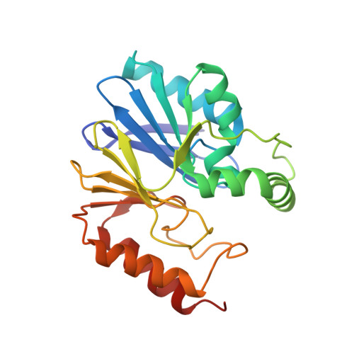
Crystal Structure of the Di-Zinc Carbapenemase CphA from Aeromonas Hydrophila


Experimental Data Snapshot

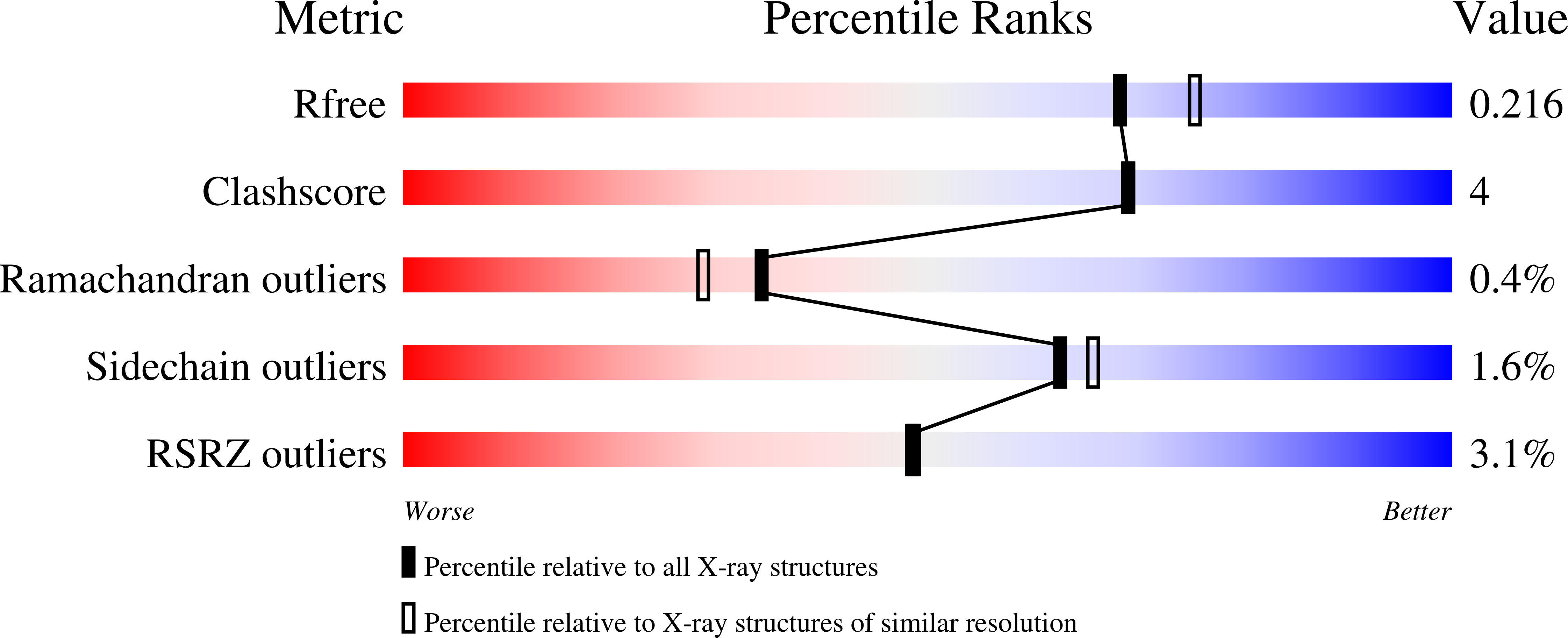
  • Method: X-RAY DIFFRACTION
  • Resolution: 2.03 Å
  • R-Value Free: 0.202 
  • R-Value Work: 0.157 
  • R-Value Observed: 0.159 

Starting Model: experimental
View more details

wwPDB Validation   3D Report Full Report


This is version 1.2 of the entry. See complete history


Literature

The structure of the dizinc subclass B2 metallo-beta-lactamase CphA reveals that the second inhibitory zinc ion binds in the histidine site

Bebrone, C.Delbruck, H.Kupper, M.B.Schlomer, P.Willmann, C.Frere, J.-M.Fischer, R.Galleni, M.Hoffmann, K.M.V.

(2009) Antimicrob Agents Chemother 53: 4464-4471

  • DOI: https://doi.org/10.1128/AAC.00288-09
  • Primary Citation of Related Structures:  
    3F9O, 3FAI

  • PubMed Abstract: 

    Bacteria can defend themselves against beta-lactam antibiotics through the expression of class B beta-lactamases, which cleave the beta-lactam amide bond and render the molecule harmless. There are three subclasses of class B beta-lactamases (B1, B2, and B3), all of which require Zn2+ for activity and can bind either one or two zinc ions. Whereas the B1 and B3 metallo-beta-lactamases are most active as dizinc enzymes, subclass B2 enzymes, such as Aeromonas hydrophila CphA, are inhibited by the binding of a second zinc ion. We crystallized A. hydrophila CphA in order to determine the binding site of the inhibitory zinc ion. X-ray data from zinc-saturated crystals allowed us to solve the crystal structures of the dizinc forms of the wild-type enzyme and N220G mutant. The first zinc ion binds in the cysteine site, as previously determined for the monozinc form of the enzyme. The second zinc ion occupies a slightly modified histidine site, where the conserved His118 and His196 residues act as metal ligands. This atypical coordination sphere probably explains the rather high dissociation constant for the second zinc ion compared to those observed with enzymes of subclasses B1 and B3. Inhibition by the second zinc ion results from immobilization of the catalytically important His118 and His196 residues, as well as the folding of the Gly232-Asn233 loop into a position that covers the active site.


  • Organizational Affiliation

    Centre for Protein Engineering, University of Liège, Allée du 6 Août B6, Sart-Tilman, 4000 Liège, Belgium. carine.bebrone@ulg.ac.be


Macromolecules
Find similar proteins by:  (by identity cutoff)  |  3D Structure
Entity ID: 1
MoleculeChains Sequence LengthOrganismDetailsImage
Beta-lactamase227Aeromonas hydrophilaMutation(s): 0 
Gene Names: cphA
EC: 3.5.2.6
UniProt
Find proteins for P26918 (Aeromonas hydrophila)
Explore P26918 
Go to UniProtKB:  P26918
Entity Groups  
Sequence Clusters30% Identity50% Identity70% Identity90% Identity95% Identity100% Identity
UniProt GroupP26918
Sequence Annotations
Expand
  • Reference Sequence
Small Molecules
Ligands 4 Unique
IDChains Name / Formula / InChI Key2D Diagram3D Interactions
SO4
Query on SO4

Download Ideal Coordinates CCD File 
B [auth A],
C [auth A],
D [auth A],
E [auth A]
SULFATE ION
O4 S
QAOWNCQODCNURD-UHFFFAOYSA-L
ZN
Query on ZN

Download Ideal Coordinates CCD File 
N [auth A],
O [auth A],
P [auth A]
ZINC ION
Zn
PTFCDOFLOPIGGS-UHFFFAOYSA-N
CO3
Query on CO3

Download Ideal Coordinates CCD File 
F [auth A],
G [auth A]
CARBONATE ION
C O3
BVKZGUZCCUSVTD-UHFFFAOYSA-L
CL
Query on CL

Download Ideal Coordinates CCD File 
H [auth A]
I [auth A]
J [auth A]
K [auth A]
L [auth A]
H [auth A],
I [auth A],
J [auth A],
K [auth A],
L [auth A],
M [auth A]
CHLORIDE ION
Cl
VEXZGXHMUGYJMC-UHFFFAOYSA-M
Experimental Data & Validation

Experimental Data

  • Method: X-RAY DIFFRACTION
  • Resolution: 2.03 Å
  • R-Value Free: 0.202 
  • R-Value Work: 0.157 
  • R-Value Observed: 0.159 
  • Space Group: C 2 2 21
Unit Cell:
Length ( Å )Angle ( ˚ )
a = 42.8α = 90
b = 101.05β = 90
c = 116.49γ = 90
Software Package:
Software NamePurpose
MAR345dtbdata collection
MOLREPphasing
REFMACrefinement
XDSdata reduction
SCALAdata scaling

Structure Validation

View Full Validation Report


Entry History 

Deposition Data

Revision History  (Full details and data files)

  • Version 1.0: 2009-09-01
    Type: Initial release
  • Version 1.1: 2011-07-13
    Changes: Advisory, Version format compliance
  • Version 1.2: 2023-11-01
    Changes: Data collection, Database references, Derived calculations, Refinement description