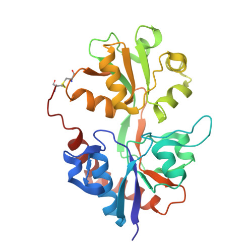
3FAT

X-ray structure of iGluR4 flip ligand-binding core (S1S2) in complex with (S)-AMPA at 1.90A resolution


Experimental Data Snapshot

  • Method: X-RAY DIFFRACTION
  • Resolution: 1.90 Å
  • R-Value Free: 0.216 
  • R-Value Work: 0.175 
  • R-Value Observed: 0.177 

Starting Model: experimental
View more details

wwPDB Validation   3D Report Full Report


Ligand Structure Quality Assessment 


This is version 1.4 of the entry. See complete history


Literature

Molecular mechanism of agonist recognition by the ligand-binding core of the ionotropic glutamate receptor 4

Kasper, C.Frydenvang, K.Naur, P.Gajhede, M.Pickering, D.S.Kastrup, J.S.

(2008) FEBS Lett 582: 4089-4094

  • DOI: https://doi.org/10.1016/j.febslet.2008.11.005
  • Primary Citation of Related Structures:  
    3FAS, 3FAT

  • PubMed Abstract: 

    The alpha-amino-3-hydroxy-5-methyl-4-isoxazolepropionic acid (AMPA) class of ionotropic glutamate receptors comprises four different subunits: iGluR1/iGluR2 and iGluR3/iGluR4 forming two subgroups. Three-dimensional structures have been reported only of the ligand-binding core of iGluR2. Here, we present two X-ray structures of a soluble construct of the R/G unedited flip splice variant of the ligand-binding core of iGluR4 (iGluR4(i)(R)-S1S2) in complex with glutamate or AMPA. Subtle, but important differences are found in the ligand-binding cavity between the two AMPA receptor subgroups at position 724 (Tyr in iGluR1/iGluR2 and Phe in iGluR3/iGluR4), which in iGluR4 may lead to displacement of a water molecule and hence points to the possibility to make subgroup specific ligands.


  • Organizational Affiliation

    Department of Medicinal Chemistry, Faculty of Pharmaceutical Sciences, University of Copenhagen, Universitetsparken 2, DK-2100 Copenhagen, Denmark.


Macromolecules
Find similar proteins by:  (by identity cutoff)  |  3D Structure
Entity ID: 1
MoleculeChains Sequence LengthOrganismDetailsImage
Glutamate receptor 4
A, B, C
260Rattus norvegicusMutation(s): 0 
UniProt
Find proteins for P19493 (Rattus norvegicus)
Explore P19493 
Go to UniProtKB:  P19493
Entity Groups  
Sequence Clusters30% Identity50% Identity70% Identity90% Identity95% Identity100% Identity
UniProt GroupP19493
Sequence Annotations
Expand
  • Reference Sequence
Small Molecules
Ligands 4 Unique
IDChains Name / Formula / InChI Key2D Diagram3D Interactions
AMQ
Query on AMQ

Download Ideal Coordinates CCD File 
D [auth A],
K [auth B],
T [auth C]
(S)-ALPHA-AMINO-3-HYDROXY-5-METHYL-4-ISOXAZOLEPROPIONIC ACID
C7 H10 N2 O4
UUDAMDVQRQNNHZ-YFKPBYRVSA-N
SO4
Query on SO4

Download Ideal Coordinates CCD File 
E [auth A]
F [auth A]
G [auth A]
H [auth A]
L [auth B]
E [auth A],
F [auth A],
G [auth A],
H [auth A],
L [auth B],
U [auth C],
V [auth C]
SULFATE ION
O4 S
QAOWNCQODCNURD-UHFFFAOYSA-L
GOL
Query on GOL

Download Ideal Coordinates CCD File 
I [auth A]
M [auth B]
N [auth B]
O [auth B]
P [auth B]
I [auth A],
M [auth B],
N [auth B],
O [auth B],
P [auth B],
W [auth C],
X [auth C],
Y [auth C],
Z [auth C]
GLYCEROL
C3 H8 O3
PEDCQBHIVMGVHV-UHFFFAOYSA-N
ACY
Query on ACY

Download Ideal Coordinates CCD File 
AA [auth C]
BA [auth C]
J [auth A]
Q [auth B]
R [auth B]
AA [auth C],
BA [auth C],
J [auth A],
Q [auth B],
R [auth B],
S [auth B]
ACETIC ACID
C2 H4 O2
QTBSBXVTEAMEQO-UHFFFAOYSA-N
Binding Affinity Annotations 
IDSourceBinding Affinity
AMQ PDBBind:  3FAT Kd: 22.8 (nM) from 1 assay(s)
Experimental Data & Validation

Experimental Data

  • Method: X-RAY DIFFRACTION
  • Resolution: 1.90 Å
  • R-Value Free: 0.216 
  • R-Value Work: 0.175 
  • R-Value Observed: 0.177 
  • Space Group: C 1 2 1
Unit Cell:
Length ( Å )Angle ( ˚ )
a = 105.175α = 90
b = 169.531β = 120.58
c = 73.883γ = 90
Software Package:
Software NamePurpose
SCALAdata scaling
PHASERphasing
PHENIXrefinement
PDB_EXTRACTdata extraction
MAR345dtbdata collection
MOSFLMdata reduction

Structure Validation

View Full Validation Report


Ligand Structure Quality Assessment 


Entry History 

Deposition Data

Revision History  (Full details and data files)

  • Version 1.0: 2008-12-09
    Type: Initial release
  • Version 1.1: 2011-07-13
    Changes: Non-polymer description, Version format compliance
  • Version 1.2: 2017-08-09
    Changes: Data collection, Refinement description, Source and taxonomy
  • Version 1.3: 2023-11-01
    Changes: Data collection, Database references, Derived calculations, Refinement description
  • Version 1.4: 2024-10-16
    Changes: Structure summary