3FED | pdb_00003fed

The high resolution structure of human glutamate carboxypeptidase III (GCPIII/NAALADase II) in complex with a transition state analog of Glu-Glu


Experimental Data Snapshot

  • Method: X-RAY DIFFRACTION
  • Resolution: 1.29 Å
  • R-Value Free: 
    0.147 (Depositor), 0.146 (DCC) 
  • R-Value Work: 
    0.131 (Depositor), 0.140 (DCC) 
  • R-Value Observed: 
    0.131 (Depositor) 

Starting Model: experimental
View more details

wwPDB Validation 3D Report Full Report

Validation slider image for 3FED

Ligand Structure Quality Assessment 


This is version 2.2 of the entry. See complete history

Literature

Structural insight into the evolutionary and pharmacologic homology of glutamate carboxypeptidases II and III

Hlouchova, K.Barinka, C.Konvalinka, J.Lubkowski, J.

(2009) FEBS J 276: 4448-4462

  • DOI: https://doi.org/10.1111/j.1742-4658.2009.07152.x
  • Primary Citation Related Structures: 
    3FEC, 3FED, 3FEE, 3FF3

  • PubMed Abstract: 

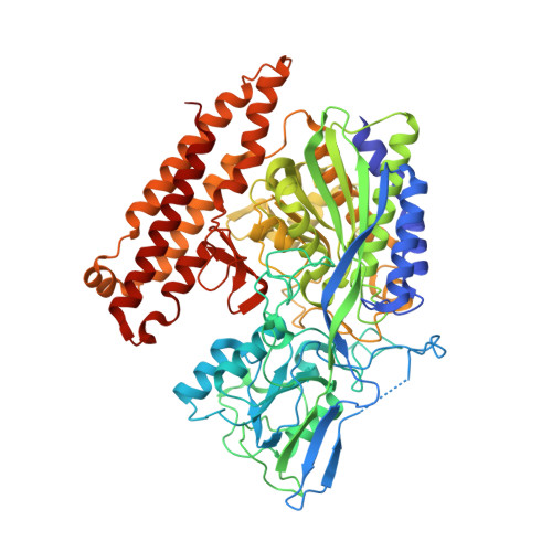
    Glutamate carboxypeptidase III (GCPIII) is a metalloenzyme that belongs to the transferrin receptor/glutamate carboxypeptidase II (GCPII; EC 3.4.17.21) superfamily. GCPIII has been studied mainly because of its evolutionary relationship to GCPII, an enzyme involved in a variety of neuropathologies and malignancies, such as glutamatergic neurotoxicity and prostate cancer. Given the potential functional and pharmacological overlap between GCPIII and GCPII, studies addressing the structural and physiological properties of GCPIII are crucial for obtaining a deeper understanding of the GCPII/GCPIII system. In the present study, we report high-resolution crystal structures of the human GCPIII ectodomain in a 'pseudo-unliganded' state and in a complex with: (a) L-glutamate (a product of hydrolysis); (b) a phosphapeptide transition state mimetic, namely (2S,3'S)-{[(3'-amino-3'-carboxy-propyl)-hydroxyphosphinoyl]methyl}-pentanedioic acid; and (c) quisqualic acid, a glutamate biostere. Our data reveal the overall fold and quaternary arrangement of the GCPIII molecule, define the architecture of the GCPIII substrate-binding cavity, and offer an experimental evidence for the presence of Zn(2+) ions in the bimetallic active site. Furthermore, the structures allow us to detail interactions between the enzyme and its ligands and to characterize the functional flexibility of GCPIII, which is essential for substrate recognition. A comparison of these GCPIII structures with the equivalent GCPII complexes reveals differences in the organization of specificity pockets, in surface charge distribution, and in the occupancy of the co-catalytic zinc sites. The data presented here provide information that should prove to be essential for the structurally-aided design of GCPIII-specific inhibitors and might comprise guidelines for future comparative GCPII/GCPIII studies.


  • Organizational Affiliation
    • Gilead Sciences and IOCB Research Centre, Institute of Organic Chemistry and Biochemistry, Academy of Sciences of the Czech Republic, Prague, Czech Republic.

Macromolecule Content 

  • Total Structure Weight: 81.55 kDa 
  • Atom Count: 6,774 
  • Modeled Residue Count: 690 
  • Deposited Residue Count: 707 
  • Unique protein chains: 1

Macromolecules

Find similar proteins by:|  3D Structure
Entity ID: 1
MoleculeChains  Sequence LengthOrganismDetailsImage
Glutamate carboxypeptidase III707Homo sapiensMutation(s): 0 
Gene Names: NAALAD2
EC: 3.4.17.21
UniProt & NIH Common Fund Data Resources
Find proteins for Q9Y3Q0 (Homo sapiens)
Explore Q9Y3Q0 
Go to UniProtKB:  Q9Y3Q0
PHAROS:  Q9Y3Q0
GTEx:  ENSG00000077616 
Entity Groups
Sequence Clusters30% Identity50% Identity70% Identity90% Identity95% Identity100% Identity
UniProt GroupQ9Y3Q0
Glycosylation
Glycosylation Sites: 4Go to GlyGen: Q9Y3Q0-1
Sequence Annotations
Expand
Reference Sequence

Oligosaccharides

Help  
Entity ID: 2
MoleculeChains Length2D Diagram GlycosylationD Interactions
2-acetamido-2-deoxy-beta-D-glucopyranose-(1-4)-2-acetamido-2-deoxy-beta-D-glucopyranose
B
2N-Glycosylation
Glycosylation Resources
GlyTouCan: G42666HT
GlyCosmos: G42666HT
GlyGen: G42666HT

Small Molecules

Ligands 5 Unique
IDChains Name / Formula / InChI Key2D Diagram3D Interactions
BIX

Query on BIX


Download:Ideal Coordinates CCD File
J [auth A](2S)-2-{[(S)-[(3S)-3-amino-3-carboxypropyl](hydroxy)phosphoryl]methyl}pentanedioic acid
C10 H18 N O8 P
YLHAQDFYEKJARV-RQJHMYQMSA-N
NAG

Query on NAG


Download:Ideal Coordinates CCD File
C [auth A],
D [auth A],
E [auth A]
2-acetamido-2-deoxy-beta-D-glucopyranose
C8 H15 N O6
OVRNDRQMDRJTHS-FMDGEEDCSA-N
ZN

Query on ZN


Download:Ideal Coordinates CCD File
F [auth A],
G [auth A]
ZINC ION
Zn
PTFCDOFLOPIGGS-UHFFFAOYSA-N
CA

Query on CA


Download:Ideal Coordinates CCD File
I [auth A]CALCIUM ION
Ca
BHPQYMZQTOCNFJ-UHFFFAOYSA-N
CL

Query on CL


Download:Ideal Coordinates CCD File
H [auth A]CHLORIDE ION
Cl
VEXZGXHMUGYJMC-UHFFFAOYSA-M

Experimental Data & Validation

Experimental Data

  • Method: X-RAY DIFFRACTION
  • Resolution: 1.29 Å
  • R-Value Free:  0.147 (Depositor), 0.146 (DCC) 
  • R-Value Work:  0.131 (Depositor), 0.140 (DCC) 
  • R-Value Observed: 0.131 (Depositor) 
Space Group: C 1 2 1
Unit Cell:
Length ( Å )Angle ( ˚ )
a = 122.731α = 90
b = 104.086β = 108.19
c = 77.634γ = 90
Software Package:
Software NamePurpose
MAR345dtbdata collection
Cootmodel building
REFMACrefinement
HKL-2000data reduction
HKL-2000data scaling

Structure Validation

View Full Validation Report


Ligand Structure Quality Assessment 


Entry History 

Deposition Data

Revision History  (Full details and data files)

  • Version 1.0: 2009-08-25
    Type: Initial release
  • Version 1.1: 2011-07-13
    Changes: Non-polymer description, Version format compliance
  • Version 2.0: 2020-07-29
    Type: Remediation
    Reason: Carbohydrate remediation
    Changes: Atomic model, Data collection, Database references, Derived calculations, Structure summary
  • Version 2.1: 2023-09-06
    Changes: Data collection, Database references, Refinement description, Structure summary
  • Version 2.2: 2024-10-30
    Changes: Structure summary