3IYW | pdb_00003iyw

West Nile virus in complex with Fab fragments of MAb CR4354 (fitted coordinates of envelope proteins and Fab fragments of one icosahedral ASU)


Experimental Data Snapshot

  • Method: ELECTRON MICROSCOPY
  • Resolution: 13.7 Å
  • Aggregation State: PARTICLE 
  • Reconstruction Method: SINGLE PARTICLE 

Starting Models: experimental
View more details

wwPDB Validation 3D Report Full Report

Validation slider image for 3IYW

This is version 4.1 of the entry. See complete history

Literature

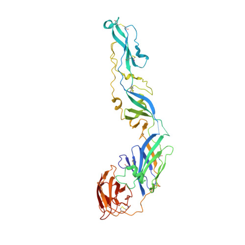
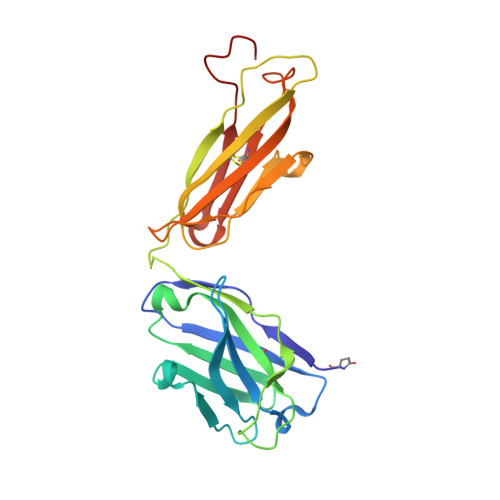
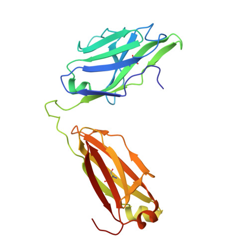
Neutralization of West Nile virus by cross-linking of its surface proteins with Fab fragments of the human monoclonal antibody CR4354.

Kaufmann, B.Vogt, M.R.Goudsmit, J.Holdaway, H.A.Aksyuk, A.A.Chipman, P.R.Kuhn, R.J.Diamond, M.S.Rossmann, M.G.

(2010) Proc Natl Acad Sci U S A 107: 18950-18955

  • DOI: https://doi.org/10.1073/pnas.1011036107
  • Primary Citation Related Structures: 
    3IYW, 3N9G

  • PubMed Abstract: 

    Many flaviviruses are significant human pathogens, with the humoral immune response playing an essential role in restricting infection and disease. CR4354, a human monoclonal antibody isolated from a patient, neutralizes West Nile virus (WNV) infection at a postattachment stage in the viral life-cycle. Here, we determined the structure of WNV complexed with Fab fragments of CR4354 using cryoelectron microscopy. The outer glycoprotein shell of a mature WNV particle is formed by 30 rafts of three homodimers of the viral surface protein E. CR4354 binds to a discontinuous epitope formed by protein segments from two neighboring E molecules, but does not cause any detectable structural disturbance on the viral surface. The epitope occurs at two independent positions within an icosahedral asymmetric unit, resulting in 120 binding sites on the viral surface. The cross-linking of the six E monomers within one raft by four CR4354 Fab fragments suggests that the antibody neutralizes WNV by blocking the pH-induced rearrangement of the E protein required for virus fusion with the endosomal membrane.


  • Organizational Affiliation
    • Department of Biological Sciences, Purdue University, West Lafayette, IN 47907-2054, USA.

Macromolecule Content 

  • Total Structure Weight: 226.84 kDa 
  • Atom Count: 16,202 
  • Modeled Residue Count: 2,100 
  • Deposited Residue Count: 2,100 
  • Unique protein chains: 3

Macromolecules

Find similar proteins by:|  3D Structure
Entity ID: 1
MoleculeChains  Sequence LengthOrganismDetailsImage
Envelope glycoprotein
A, B, C
400West Nile virusMutation(s): 0 
Membrane Entity: Yes 
UniProt
Find proteins for Q91R02 (West Nile virus)
Explore Q91R02 
Go to UniProtKB:  Q91R02
Entity Groups
Sequence Clusters30% Identity50% Identity70% Identity90% Identity95% Identity100% Identity
UniProt GroupQ91R02
Sequence Annotations
Expand
Reference Sequence
Find similar proteins by:|  3D Structure
Entity ID: 2
MoleculeChains  Sequence LengthOrganismDetailsImage
CR4354 Fab fragment X1, heavy chain HD [auth H],
F [auth K]
230Homo sapiensMutation(s): 0 
Find similar proteins by:|  3D Structure
Entity ID: 3
MoleculeChains  Sequence LengthOrganismDetailsImage
CR4354 Fab fragment X1, light chain LE [auth L],
G [auth M]
220Homo sapiensMutation(s): 0 

Oligosaccharides

Help  
Entity ID: 4
MoleculeChains Length2D Diagram GlycosylationD Interactions
2-acetamido-2-deoxy-alpha-D-glucopyranose-(1-4)-[beta-L-fucopyranose-(1-6)]2-acetamido-2-deoxy-alpha-D-glucopyranoseH [auth D],
I [auth E],
J [auth F]
3N/A
Glycosylation Resources
GlyTouCan: G24285GM
GlyCosmos: G24285GM
GlyGen: G24285GM

Small Molecules

Modified Residues  1 Unique
IDChains TypeFormula2D DiagramParent
PCA
Query on PCA
D [auth H],
F [auth K]
L-PEPTIDE LINKINGC5 H7 N O3GLN

Experimental Data & Validation

Experimental Data

  • Method: ELECTRON MICROSCOPY
  • Resolution: 13.7 Å
  • Aggregation State: PARTICLE 
  • Reconstruction Method: SINGLE PARTICLE 
EM Software:
TaskSoftware PackageVersion
RECONSTRUCTIONAuto3DEM

Structure Validation

View Full Validation Report


Entry History 

Deposition Data

Revision History  (Full details and data files)

  • Version 1.0: 2010-11-24
    Type: Initial release
  • Version 1.1: 2011-07-13
    Changes: Version format compliance
  • Version 1.2: 2018-07-18
    Changes: Data collection
  • Version 2.0: 2019-12-25
    Changes: Database references, Derived calculations, Polymer sequence
  • Version 3.0: 2020-07-29
    Type: Remediation
    Reason: Carbohydrate remediation
    Changes: Advisory, Atomic model, Data collection, Derived calculations, Structure summary
  • Version 4.0: 2024-03-27
    Changes: Advisory, Data collection, Database references, Derived calculations, Polymer sequence, Refinement description, Source and taxonomy, Structure summary
  • Version 4.1: 2024-11-20
    Changes: Structure summary