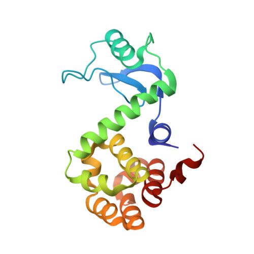
Sequential reorganization of beta-sheet topology by insertion of a single strand.
Sagermann, M., Baase, W.A., Matthews, B.W.(2006) Protein Sci 15: 1085-1092
- PubMed: 16597830
- DOI: https://doi.org/10.1110/ps.052018006
- Primary Citation of Related Structures:
2B7X, 3JR6 - PubMed Abstract:
Insertions, duplications, and deletions of sequence segments are thought to be major evolutionary mechanisms that increase the structural and functional diversity of proteins. Alternative splicing, for example, is an intracellular editing mechanism that is thought to generate isoforms for 30%-50% of all human genes. Whereas the inserted sequences usually display only minor structural rearrangements at the insertion site, recent observations indicate that they may also cause more dramatic structural displacements of adjacent structures. In the present study we test how artificially inserted sequences change the structure of the beta-sheet region in T4 lysozyme. Copies of two different beta-strands were inserted into two different loops of the beta-sheet, and the structures were determined. Not surprisingly, one insert "loops out" at its insertion site and forms a new small beta-hairpin structure. Unexpectedly, however, the second insertion leads to displacement of adjacent strands and a sequential reorganization of the beta-sheet topology. Even though the insertions were performed at two different sites, looping out occurred at the C-terminal end of the same beta-strand. Reasons as to why a non-native sequence would be recruited to replace that which occurs in the native protein are discussed. Our results illustrate how sequence insertions can facilitate protein evolution through both local and nonlocal changes in structure.
Organizational Affiliation:
Institute of Molecular Biology, Howard Hughes Medical Institute, and Department of Physics, University of Oregon, Eugene, Oregon 97403-1229, USA.


