3KYM | pdb_00003kym

Crystal structure of Li33 IgG2 di-Fab


Experimental Data Snapshot

  • Method: X-RAY DIFFRACTION
  • Resolution: 2.62 Å
  • R-Value Free: 
    0.339 (Depositor), 0.332 (DCC) 
  • R-Value Work: 
    0.259 (Depositor), 0.256 (DCC) 
  • R-Value Observed: 
    0.267 (Depositor) 

Starting Model: experimental
View more details

wwPDB Validation 3D Report Full Report

Validation slider image for 3KYM

This is version 1.3 of the entry. See complete history

Literature

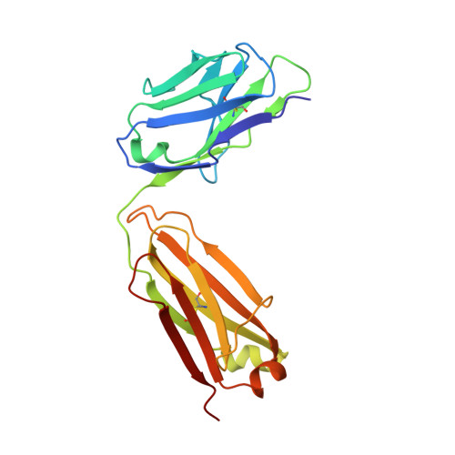
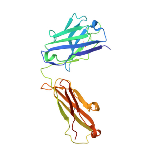
Improving the solubility of anti-LINGO-1 monoclonal antibody Li33 by isotype switching and targeted mutagenesis.

Pepinsky, R.B.Silvian, L.Berkowitz, S.A.Farrington, G.Lugovskoy, A.Walus, L.Eldredge, J.Capili, A.Mi, S.Graff, C.Garber, E.

(2010) Protein Sci 19: 954-966

  • DOI: https://doi.org/10.1002/pro.372
  • Primary Citation Related Structures: 
    3KYK, 3KYM

  • PubMed Abstract: 

    Monoclonal antibodies (Mabs) are a favorite drug platform of the biopharmaceutical industry. Currently, over 20 Mabs have been approved and several hundred others are in clinical trials. The anti-LINGO-1 Mab Li33 was selected from a large panel of antibodies by Fab phage display technology based on its extraordinary biological activity in promoting oligodendrocyte differentiation and myelination in vitro and in animal models of remyelination. However, the Li33 Fab had poor solubility when converted into a full antibody in an immunoglobulin G1 framework. A detailed analysis of the biochemical and structural features of the antibody revealed several possible reasons for its propensity to aggregate. Here, we successfully applied three molecular approaches (isotype switching, targeted mutagenesis of complementarity determining region residues, and glycosylation site insertion mutagenesis) to address the solubility problem. Through these efforts we were able to improve the solubility of the Li33 Mab from 0.3 mg/mL to >50 mg/mL and reduce aggregation to an acceptable level. These strategies can be readily applied to other proteins with solubility issues.


  • Organizational Affiliation
    • Department of Drug Discovery, Biogen Idec, Inc., Cambridge, Massachusetts 02142, USA. blake.pepinsky@biogenidec.com

Macromolecule Content 

  • Total Structure Weight: 383.29 kDa 
  • Atom Count: 26,358 
  • Modeled Residue Count: 3,449 
  • Deposited Residue Count: 3,528 
  • Unique protein chains: 2

Macromolecules

Find similar proteins by:|  3D Structure
Entity ID: 1
MoleculeChains  Sequence LengthOrganismDetailsImage
Light Chain Li33 IgG2
A, C, E, G, I
A, C, E, G, I, K, M, O
214Homo sapiensMutation(s): 0 
Find similar proteins by:|  3D Structure
Entity ID: 2
MoleculeChains  Sequence LengthOrganismDetailsImage
Heavy Chain Li33 IgG2
B, D, F, H, J
B, D, F, H, J, L, N, P
227Homo sapiensMutation(s): 0 

Experimental Data & Validation

Experimental Data

  • Method: X-RAY DIFFRACTION
  • Resolution: 2.62 Å
  • R-Value Free:  0.339 (Depositor), 0.332 (DCC) 
  • R-Value Work:  0.259 (Depositor), 0.256 (DCC) 
  • R-Value Observed: 0.267 (Depositor) 
Space Group: P 1
Unit Cell:
Length ( Å )Angle ( ˚ )
a = 91.669α = 61.46
b = 109.545β = 79.29
c = 118.433γ = 87.59
Software Package:
Software NamePurpose
HKL-2000data collection
PHASERphasing
REFMACrefinement
HKL-2000data reduction
HKL-2000data scaling

Structure Validation

View Full Validation Report


Entry History 

Deposition Data

Revision History  (Full details and data files)

  • Version 1.0: 2010-03-16
    Type: Initial release
  • Version 1.1: 2011-07-13
    Changes: Version format compliance
  • Version 1.2: 2023-09-06
    Changes: Data collection, Database references, Refinement description
  • Version 1.3: 2024-10-16
    Changes: Structure summary