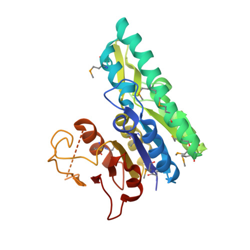
3LL5

Crystal structure of T. acidophilum isopentenyl phosphate kinase product complex


Experimental Data Snapshot

  • Method: X-RAY DIFFRACTION
  • Resolution: 1.99 Å
  • R-Value Free: 0.238 
  • R-Value Work: 0.193 
  • R-Value Observed: 0.195 

Starting Model: experimental
View more details

wwPDB Validation   3D Report Full Report


Ligand Structure Quality Assessment 


This is version 1.3 of the entry. See complete history


Literature

X-ray structures of isopentenyl phosphate kinase.

Mabanglo, M.F.Schubert, H.L.Chen, M.Hill, C.P.Poulter, C.D.

(2010) ACS Chem Biol 5: 517-527

  • DOI: https://doi.org/10.1021/cb100032g
  • Primary Citation of Related Structures:  
    3LKK, 3LL5, 3LL9

  • PubMed Abstract: 

    Isoprenoid compounds are ubiquitous in nature, participating in important biological phenomena such as signal transduction, aerobic cellular respiration, photosynthesis, insect communication, and many others. They are derived from the 5-carbon isoprenoid substrates isopentenyl diphosphate (IPP) and its isomer dimethylallyl diphosphate (DMAPP). In Archaea and Eukarya, these building blocks are synthesized via the mevalonate pathway. However, the genes required to convert mevalonate phosphate (MP) to IPP are missing in several species of Archaea. An enzyme with isopentenyl phosphate kinase (IPK) activity was recently discovered in Methanocaldococcus jannaschii (MJ), suggesting a departure from the classical sequence of converting MP to IPP. We have determined the high-resolution crystal structures of isopentenyl phosphate kinases in complex with both substrates and products from Thermoplasma acidophilum (THA), as well as the IPK from Methanothermobacter thermautotrophicus (MTH), by means of single-wavelength anomalous diffraction (SAD) and molecular replacement. A histidine residue (His50) in THA IPK makes a hydrogen bond with the terminal phosphates of IP and IPP, poising these molecules for phosphoryl transfer through an in-line geometry. Moreover, a lysine residue (Lys14) makes hydrogen bonds with nonbridging oxygen atoms at P(alpha) and P(gamma) and with the P(beta)-P(gamma) bridging oxygen atom in ATP. These interactions suggest a transition-state-stabilizing role for this residue. Lys14 is a part of a newly discovered "lysine triangle" catalytic motif in IPKs that also includes Lys5 and Lys205. Moreover, His50, Lys5, Lys14, and Lys205 are conserved in all IPKs and can therefore serve as fingerprints for identifying new homologues.


  • Organizational Affiliation

    Department of Chemistry, University of Utah, Salt Lake City, 84112, USA.


Macromolecules
Find similar proteins by:  (by identity cutoff)  |  3D Structure
Entity ID: 1
MoleculeChains Sequence LengthOrganismDetailsImage
Gamma-glutamyl kinase related protein
A, B, C, D
249Thermoplasma acidophilum DSM 1728Mutation(s): 0 
Gene Names: Ta0103
EC: 2.7.4.26
UniProt
Find proteins for Q9HLX1 (Thermoplasma acidophilum (strain ATCC 25905 / DSM 1728 / JCM 9062 / NBRC 15155 / AMRC-C165))
Explore Q9HLX1 
Go to UniProtKB:  Q9HLX1
Entity Groups  
Sequence Clusters30% Identity50% Identity70% Identity90% Identity95% Identity100% Identity
UniProt GroupQ9HLX1
Sequence Annotations
Expand
  • Reference Sequence
Small Molecules
Ligands 4 Unique
IDChains Name / Formula / InChI Key2D Diagram3D Interactions
ATP
Query on ATP

Download Ideal Coordinates CCD File 
I [auth C],
K [auth D]
ADENOSINE-5'-TRIPHOSPHATE
C10 H16 N5 O13 P3
ZKHQWZAMYRWXGA-KQYNXXCUSA-N
ADP
Query on ADP

Download Ideal Coordinates CCD File 
E [auth A],
G [auth B]
ADENOSINE-5'-DIPHOSPHATE
C10 H15 N5 O10 P2
XTWYTFMLZFPYCI-KQYNXXCUSA-N
IPE
Query on IPE

Download Ideal Coordinates CCD File 
F [auth A],
H [auth B]
3-METHYLBUT-3-ENYL TRIHYDROGEN DIPHOSPHATE
C5 H12 O7 P2
NUHSROFQTUXZQQ-UHFFFAOYSA-N
IP8
Query on IP8

Download Ideal Coordinates CCD File 
J [auth C],
L [auth D]
Isopentenyl phosphate
C5 H11 O4 P
QMZRXYCCCYYMHF-UHFFFAOYSA-N
Modified Residues  1 Unique
IDChains TypeFormula2D DiagramParent
MSE
Query on MSE
A, B, C, D
L-PEPTIDE LINKINGC5 H11 N O2 SeMET
Experimental Data & Validation

Experimental Data

  • Method: X-RAY DIFFRACTION
  • Resolution: 1.99 Å
  • R-Value Free: 0.238 
  • R-Value Work: 0.193 
  • R-Value Observed: 0.195 
  • Space Group: C 1 2 1
Unit Cell:
Length ( Å )Angle ( ˚ )
a = 187.104α = 90
b = 42.787β = 113.19
c = 134.895γ = 90
Software Package:
Software NamePurpose
CrystalCleardata collection
PHASERphasing
PHENIXrefinement
HKL-2000data reduction
HKL-2000data scaling

Structure Validation

View Full Validation Report


Ligand Structure Quality Assessment 


Entry History 

Deposition Data

Revision History  (Full details and data files)

  • Version 1.0: 2010-06-02
    Type: Initial release
  • Version 1.1: 2011-07-13
    Changes: Version format compliance
  • Version 1.2: 2024-04-03
    Changes: Data collection, Database references, Derived calculations, Refinement description
  • Version 1.3: 2024-11-27
    Changes: Structure summary