3LZO | pdb_00003lzo

Crystal Structure Analysis of the copper-reconstituted P19 protein from Campylobacter jejuni at 1.65 A at pH 10.0


Experimental Data Snapshot

  • Method: X-RAY DIFFRACTION
  • Resolution: 1.65 Å
  • R-Value Free: 
    0.187 (Depositor), 0.196 (DCC) 
  • R-Value Work: 
    0.139 (Depositor), 0.151 (DCC) 
  • R-Value Observed: 
    0.141 (Depositor) 

Starting Model: experimental
View more details

wwPDB Validation 3D Report Full Report

Validation slider image for 3LZO

This is version 1.3 of the entry. See complete history

Literature

Structure and Function of P19, a High-Affinity Iron Transporter of the Human Pathogen Campylobacter jejuni.

Chan, A.C.Doukov, T.I.Scofield, M.Tom-Yew, S.A.Ramin, A.B.Mackichan, J.K.Gaynor, E.C.Murphy, M.E.

(2010) J Mol Biology 401: 590-604

  • DOI: https://doi.org/10.1016/j.jmb.2010.06.038
  • Primary Citation Related Structures: 
    3LZL, 3LZN, 3LZO, 3LZP, 3LZQ, 3LZR

  • PubMed Abstract: 

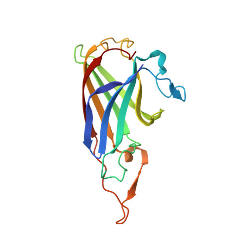
    Campylobacter jejuni, a major cause of acute bacterial diarrhea in humans, expresses numerous proteins to import diverse forms of essential iron. The expression of p19 and an adjacent iron transporter homologue (ftr1) is strongly induced upon iron limitation, suggesting a function in iron acquisition. Here, we show that the loss of P19 alone is detrimental to growth on iron-restricted media. Furthermore, metal binding analysis demonstrates that recombinant P19 has distinct copper and iron binding sites. Crystal structures of P19 have been solved to 1.41 A resolution, revealing an immunoglobulin-like fold. A P19 homodimer in which both monomers contribute ligands to two equivalent copper sites located adjacent to methionine-rich patches is observed. Copper coordination occurs via three histidine residues (His42, His95, and His132) and Met88. A solvent channel lined with conserved acidic residues leads to the copper site. Soaking crystals with a solution of manganese as iron analog reveals a second metal binding site in this solvent channel (metal-metal distance, 7.7 A). Glu44 lies between the metal sites and displays multiple conformations in the crystal structures, suggesting a role in regulating metal-metal interaction. Dimerization is shown to be metal dependent in vitro and is detected in vivo by cross-linking.


  • Organizational Affiliation
    • Department of Microbiology and Immunology, Life Sciences Institute, 2350 Health Sciences Mall, The University of British Columbia, Vancouver, BC, Canada V6T 1Z3.

Macromolecule Content 

  • Total Structure Weight: 35.5 kDa 
  • Atom Count: 2,836 
  • Modeled Residue Count: 318 
  • Deposited Residue Count: 318 
  • Unique protein chains: 1

Macromolecules

Find similar proteins by:|  3D Structure
Entity ID: 1
MoleculeChains  Sequence LengthOrganismDetailsImage
P19 protein
A, B
159Campylobacter jejuni subsp. jejuni 81-176Mutation(s): 0 
Gene Names: CJJ81176_1650Gene Cj81176_1659
UniProt
Find proteins for A0A0H3PA01 (Campylobacter jejuni subsp. jejuni serotype O:23/36 (strain 81-176))
Explore A0A0H3PA01 
Go to UniProtKB:  A0A0H3PA01
Entity Groups
Sequence Clusters30% Identity50% Identity70% Identity90% Identity95% Identity100% Identity
UniProt GroupA0A0H3PA01
Sequence Annotations
Expand
Reference Sequence

Experimental Data & Validation

Experimental Data

  • Method: X-RAY DIFFRACTION
  • Resolution: 1.65 Å
  • R-Value Free:  0.187 (Depositor), 0.196 (DCC) 
  • R-Value Work:  0.139 (Depositor), 0.151 (DCC) 
  • R-Value Observed: 0.141 (Depositor) 
Space Group: P 21 21 2
Unit Cell:
Length ( Å )Angle ( ˚ )
a = 55.831α = 90
b = 72.581β = 90
c = 78.817γ = 90
Software Package:
Software NamePurpose
Blu-Icedata collection
REFMACrefinement
XDSdata reduction
XSCALEdata scaling
REFMACphasing

Structure Validation

View Full Validation Report


Entry History 

Deposition Data

Revision History  (Full details and data files)

  • Version 1.0: 2010-07-21
    Type: Initial release
  • Version 1.1: 2011-07-13
    Changes: Version format compliance
  • Version 1.2: 2024-02-21
    Changes: Data collection, Database references, Derived calculations
  • Version 1.3: 2024-04-03
    Changes: Refinement description