3N5O | pdb_00003n5o

Crystal structure of putative glutathione transferase from Coccidioides immitis bound to glutathione


Experimental Data Snapshot

  • Method: X-RAY DIFFRACTION
  • Resolution: 1.85 Å
  • R-Value Free: 
    0.205 (Depositor), 0.203 (DCC) 
  • R-Value Work: 
    0.158 (Depositor), 0.158 (DCC) 
  • R-Value Observed: 
    0.160 (Depositor) 

Starting Model: experimental
View more details

wwPDB Validation 3D Report Full Report

Validation slider image for 3N5O

Ligand Structure Quality Assessment 


This is version 1.3 of the entry. See complete history

Literature

Structures of a putative zeta-class glutathione S-transferase from the pathogenic fungus Coccidioides immitis.

Edwards, T.E.Bryan, C.M.Leibly, D.J.Dieterich, S.H.Abendroth, J.Sankaran, B.Sivam, D.Staker, B.L.Van Voorhis, W.C.Myler, P.J.Stewart, L.J.

(2011) Acta Crystallogr Sect F Struct Biol Cryst Commun 67: 1038-1043

  • DOI: https://doi.org/10.1107/S1744309111009493
  • Primary Citation Related Structures: 
    3LG6, 3N5O

  • PubMed Abstract: 

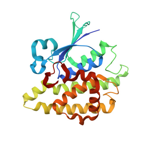
    Coccidioides immitis is a pathogenic fungus populating the southwestern United States and is a causative agent of coccidioidomycosis, sometimes referred to as Valley Fever. Although the genome of this fungus has been sequenced, many operons are not properly annotated. Crystal structures are presented for a putative uncharacterized protein that shares sequence similarity with ζ-class glutathione S-transferases (GSTs) in both apo and glutathione-bound forms. The apo structure reveals a nonsymmetric homodimer with each protomer comprising two subdomains: a C-terminal helical domain and an N-terminal thioredoxin-like domain that is common to all GSTs. Half-site binding is observed in the glutathione-bound form. Considerable movement of some components of the active site relative to the glutathione-free form was observed, indicating an induced-fit mechanism for cofactor binding. The sequence homology, structure and half-site occupancy imply that the protein is a ζ-class glutathione S-transferase, a maleylacetoacetate isomerase (MAAI).


  • Organizational Affiliation
    • Seattle Structural Genomics Center for Infectious Disease, USA. tedwards@embios.com

Macromolecule Content 

  • Total Structure Weight: 52.42 kDa 
  • Atom Count: 3,930 
  • Modeled Residue Count: 442 
  • Deposited Residue Count: 470 
  • Unique protein chains: 1

Macromolecules

Find similar proteins by:|  3D Structure
Entity ID: 1
MoleculeChains  Sequence LengthOrganismDetailsImage
glutathione transferase
A, B
235Coccidioides immitisMutation(s): 0 
Gene Names: COI
EC: 2.5.1.18
UniProt
Find proteins for D2YW48 (Coccidioides immitis (strain RS))
Explore D2YW48 
Go to UniProtKB:  D2YW48
Entity Groups
Sequence Clusters30% Identity50% Identity70% Identity90% Identity95% Identity100% Identity
UniProt GroupD2YW48
Sequence Annotations
Expand
Reference Sequence

Biologically Interesting Molecules (External Reference) 

1 Unique

Experimental Data & Validation

Experimental Data

  • Method: X-RAY DIFFRACTION
  • Resolution: 1.85 Å
  • R-Value Free:  0.205 (Depositor), 0.203 (DCC) 
  • R-Value Work:  0.158 (Depositor), 0.158 (DCC) 
  • R-Value Observed: 0.160 (Depositor) 
Space Group: C 2 2 21
Unit Cell:
Length ( Å )Angle ( ˚ )
a = 49.61α = 90
b = 110.93β = 90
c = 168.49γ = 90
Software Package:
Software NamePurpose
XSCALEdata scaling
PHASERphasing
REFMACrefinement
PDB_EXTRACTdata extraction
StructureStudiodata collection
XDSdata reduction

Structure Validation

View Full Validation Report


Ligand Structure Quality Assessment 


Entry History 

Deposition Data

Revision History  (Full details and data files)

  • Version 1.0: 2010-06-16
    Type: Initial release
  • Version 1.1: 2011-07-13
    Changes: Version format compliance
  • Version 1.2: 2011-09-21
    Changes: Database references
  • Version 1.3: 2023-09-06
    Changes: Data collection, Database references, Derived calculations, Refinement description