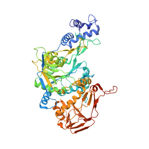
3OH1

Protein structure of USP from L. major bound to URIDINE-5'-DIPHOSPHATE-Galacturonic acid


Experimental Data Snapshot

  • Method: X-RAY DIFFRACTION
  • Resolution: 2.18 Å
  • R-Value Free: 0.243 
  • R-Value Work: 0.200 
  • R-Value Observed: 0.202 

Starting Model: experimental
View more details

wwPDB Validation   3D Report Full Report


Ligand Structure Quality Assessment 


This is version 1.2 of the entry. See complete history


Literature

Structural basis for the broad substrate range of the UDP-sugar pyrophosphorylase from Leishmania major.

Dickmanns, A.Damerow, S.Neumann, P.Schulz, E.C.Lamerz, A.C.Routier, F.H.Ficner, R.

(2011) J Mol Biol 405: 461-478

  • DOI: https://doi.org/10.1016/j.jmb.2010.10.057
  • Primary Citation of Related Structures:  
    3OGZ, 3OH0, 3OH1, 3OH2, 3OH3, 3OH4

  • PubMed Abstract: 

    Nucleotide sugars and the enzymes that are responsible for their synthesis are indispensable for the production of complex carbohydrates and, thus, for elaboration of a protective cellular coat for many organisms such as the protozoan parasite Leishmania. These activated sugars are synthesized de novo or derived from salvaged monosaccharides. In addition to UDP-glucose (UDP-Glc) pyrophosphorylase, which catalyzes the formation of UDP-Glc from substrates UTP and glucose-1-phosphate, Leishmania major and plants express a UDP-sugar pyrophosphorylase (USP) that exhibits broad substrate specificity in vitro. The enzyme, likely involved in monosaccharide salvage, preferentially generates UDP-Glc and UDP-galactose, but it may also activate other hexose- or pentose-1-phosphates such as galacturonic acid-1-phosphate or arabinose-1-phosphate. In order to gain insight into structural features governing the differences in substrate specificity, we determined the crystal structure of the L. major USP in the APO-, UTP-, and UDP-sugar-bound conformations. The overall tripartite structure of USP exhibits a significant structural homology to other nucleotidyldiphosphate-glucose pyrophosphorylases. The obtained USP structures reveal the structural rearrangements occurring during the stepwise binding process of the substrates. Moreover, the different product complexes explain the broad substrate specificity of USP, which is enabled by structural changes in the sugar binding region of the active site.


  • Organizational Affiliation

    Institut für Mikrobiologie und Genetik & GZMB, Georg-August-Universität Göttingen, Justus-von-Liebig-Weg 11, D-37077 Göttingen, Germany.


Macromolecules
Find similar proteins by:  (by identity cutoff)  |  3D Structure
Entity ID: 1
MoleculeChains Sequence LengthOrganismDetailsImage
UDP-sugar pyrophosphorylase641Leishmania majorMutation(s): 0 
Gene Names: USP
EC: 2.7.7.64
UniProt
Find proteins for D3G6S4 (Leishmania major)
Explore D3G6S4 
Go to UniProtKB:  D3G6S4
Entity Groups  
Sequence Clusters30% Identity50% Identity70% Identity90% Identity95% Identity100% Identity
UniProt GroupD3G6S4
Sequence Annotations
Expand
  • Reference Sequence
Small Molecules
Ligands 2 Unique
IDChains Name / Formula / InChI Key2D Diagram3D Interactions
UGB
Query on UGB

Download Ideal Coordinates CCD File 
B [auth A](2S,3R,4S,5R,6R)-6-[[[(2R,3S,4R,5R)-5-(2,4-dioxopyrimidin-1-yl)-3,4-dihydroxy-oxolan-2-yl]methoxy-hydroxy-phosphoryl]oxy-hydroxy-phosphoryl]oxy-3,4,5-trihydroxy-oxane-2-carboxylic acid
C15 H22 N2 O18 P2
HDYANYHVCAPMJV-GXNRKQDOSA-N
GOL
Query on GOL

Download Ideal Coordinates CCD File 
C [auth A],
D [auth A]
GLYCEROL
C3 H8 O3
PEDCQBHIVMGVHV-UHFFFAOYSA-N
Experimental Data & Validation

Experimental Data

  • Method: X-RAY DIFFRACTION
  • Resolution: 2.18 Å
  • R-Value Free: 0.243 
  • R-Value Work: 0.200 
  • R-Value Observed: 0.202 
  • Space Group: C 1 2 1
Unit Cell:
Length ( Å )Angle ( ˚ )
a = 108.226α = 90
b = 122.32β = 105.42
c = 61.221γ = 90
Software Package:
Software NamePurpose
SCALEPACKdata scaling
PHENIXrefinement
PDB_EXTRACTdata extraction
MAR345dtbdata collection
HKL-2000data reduction
CNSphasing

Structure Validation

View Full Validation Report


Ligand Structure Quality Assessment 


Entry History 

Deposition Data

Revision History  (Full details and data files)

  • Version 1.0: 2010-11-17
    Type: Initial release
  • Version 1.1: 2011-07-13
    Changes: Version format compliance
  • Version 1.2: 2023-09-06
    Changes: Data collection, Database references, Derived calculations, Refinement description