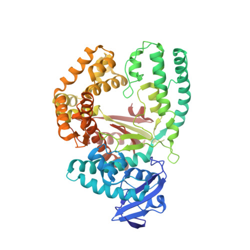
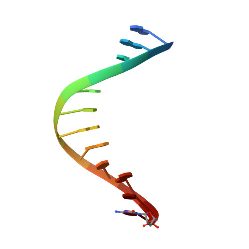
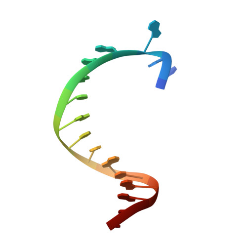
3OJS

Snapshots of the large fragment of DNA polymerase I from Thermus Aquaticus processing C5 modified thymidines


Experimental Data Snapshot

  • Method: X-RAY DIFFRACTION
  • Resolution: 1.90 Å
  • R-Value Free: 0.212 
  • R-Value Work: 0.163 
  • R-Value Observed: 0.166 

Starting Model: experimental
View more details

wwPDB Validation   3D Report Full Report


Ligand Structure Quality Assessment 


This is version 1.4 of the entry. See complete history


Literature

Structural basis for the synthesis of nucleobase modified DNA by Thermus aquaticus DNA polymerase.

Obeid, S.Baccaro, A.Welte, W.Diederichs, K.Marx, A.

(2010) Proc Natl Acad Sci U S A 107: 21327-21331

  • DOI: https://doi.org/10.1073/pnas.1013804107
  • Primary Citation of Related Structures:  
    3OJS, 3OJU

  • PubMed Abstract: 

    Numerous 2'-deoxynucleoside triphosphates (dNTPs) that are functionalized with spacious modifications such as dyes and affinity tags like biotin are substrates for DNA polymerases. They are widely employed in many cutting-edge technologies like advanced DNA sequencing approaches, microarrays, and single molecule techniques. Modifications attached to the nucleobase are accepted by many DNA polymerases, and thus, dNTPs bearing nucleobase modifications are predominantly employed. When pyrimidines are used the modifications are almost exclusively at the C5 position to avoid disturbing of Watson-Crick base pairing ability. However, the detailed molecular mechanism by which C5 modifications are processed by a DNA polymerase is poorly understood. Here, we present the first crystal structures of a DNA polymerase from Thermus aquaticus processing two C5 modified substrates that are accepted by the enzyme with different efficiencies. The structures were obtained as ternary complex of the enzyme bound to primer/template duplex with the respective modified dNTP in position poised for catalysis leading to incorporation. Thus, the study provides insights into the incorporation mechanism of the modified nucleotides elucidating how bulky modifications are accepted by the enzyme. The structures show a varied degree of perturbation of the enzyme substrate complexes depending on the nature of the modifications suggesting design principles for future developments of modified substrates for DNA polymerases.


  • Organizational Affiliation

    Department of Chemistry, Konstanz Research School Chemical Biology, University of Konstanz, Universitätsstrasse 10, D 78457 Konstanz, Germany.


Macromolecules

Find similar proteins by:  (by identity cutoff)  |  3D Structure
Entity ID: 1
MoleculeChains Sequence LengthOrganismDetailsImage
DNA polymerase I, thermostable540Thermus aquaticusMutation(s): 0 
Gene Names: polApol1
EC: 2.7.7.7
UniProt
Find proteins for P19821 (Thermus aquaticus)
Explore P19821 
Go to UniProtKB:  P19821
Entity Groups  
Sequence Clusters30% Identity50% Identity70% Identity90% Identity95% Identity100% Identity
UniProt GroupP19821
Sequence Annotations
Expand
  • Reference Sequence

Find similar nucleic acids by:  Sequence   |   3D Structure  

Entity ID: 2
MoleculeChains LengthOrganismImage
DNA (5'-D(*GP*AP*CP*CP*AP*CP*GP*GP*CP*GP*CP*(DOC))-3')12N/A
Sequence Annotations
Expand
  • Reference Sequence

Find similar nucleic acids by:  Sequence   |   3D Structure  

Entity ID: 3
MoleculeChains LengthOrganismImage
DNA (5'-D(*AP*AP*AP*AP*GP*GP*CP*GP*CP*CP*GP*TP*GP*GP*TP*C)-3')16N/A
Sequence Annotations
Expand
  • Reference Sequence
Small Molecules
Ligands 5 Unique
IDChains Name / Formula / InChI Key2D Diagram3D Interactions
XJS
Query on XJS

Download Ideal Coordinates CCD File 
D [auth A]2'-deoxy-5-[9-(3-{[4-(diethylamino)-4-oxobutanoyl]amino}propyl)-18-ethyl-5,8,14,17-tetraoxo-4,9,13,18-tetraazaicos-1-yn-1-yl]uridine 5'-(tetrahydrogen triphosphate)
C38 H63 N8 O20 P3
FRKZJUQJYOJKOB-CMUUVTEDSA-N
PGE
Query on PGE

Download Ideal Coordinates CCD File 
L [auth A],
N [auth A],
O [auth A],
S [auth C]
TRIETHYLENE GLYCOL
C6 H14 O4
ZIBGPFATKBEMQZ-UHFFFAOYSA-N
GOL
Query on GOL

Download Ideal Coordinates CCD File 
E [auth A]
F [auth A]
G [auth A]
H [auth A]
I [auth A]
E [auth A],
F [auth A],
G [auth A],
H [auth A],
I [auth A],
R [auth B],
T [auth C],
U [auth C]
GLYCEROL
C3 H8 O3
PEDCQBHIVMGVHV-UHFFFAOYSA-N
ACT
Query on ACT

Download Ideal Coordinates CCD File 
M [auth A],
P [auth A],
Q [auth A]
ACETATE ION
C2 H3 O2
QTBSBXVTEAMEQO-UHFFFAOYSA-M
MG
Query on MG

Download Ideal Coordinates CCD File 
J [auth A],
K [auth A]
MAGNESIUM ION
Mg
JLVVSXFLKOJNIY-UHFFFAOYSA-N
Experimental Data & Validation

Experimental Data

  • Method: X-RAY DIFFRACTION
  • Resolution: 1.90 Å
  • R-Value Free: 0.212 
  • R-Value Work: 0.163 
  • R-Value Observed: 0.166 
  • Space Group: P 31 2 1
Unit Cell:
Length ( Å )Angle ( ˚ )
a = 107.829α = 90
b = 107.829β = 90
c = 90.197γ = 120
Software Package:
Software NamePurpose
PHENIXmodel building
PHENIXrefinement
XDSdata reduction
XDSdata scaling
PHENIXphasing

Structure Validation

View Full Validation Report


Ligand Structure Quality Assessment 


Entry History 

Deposition Data

Revision History  (Full details and data files)

  • Version 1.0: 2010-12-15
    Type: Initial release
  • Version 1.1: 2011-07-13
    Changes: Version format compliance
  • Version 1.2: 2013-03-27
    Changes: Database references
  • Version 1.3: 2017-11-08
    Changes: Refinement description
  • Version 1.4: 2023-09-06
    Changes: Data collection, Database references, Derived calculations, Refinement description