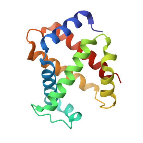
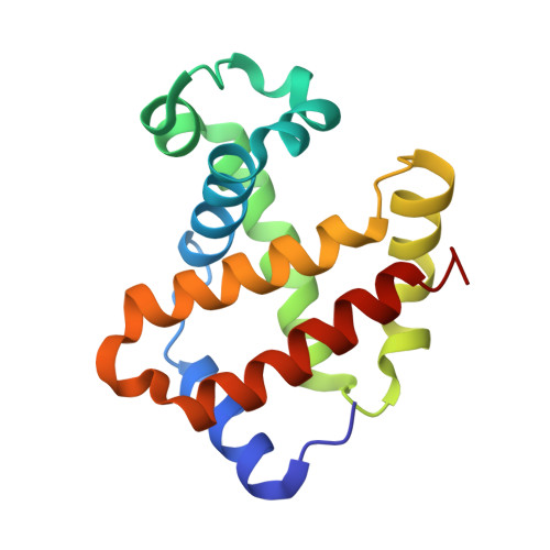
3P5Q | pdb_00003p5q

Ferric R-state human aquomethemoglobin


Experimental Data Snapshot

  • Method: X-RAY DIFFRACTION
  • Resolution: 2.00 Å
  • R-Value Free: 
    0.267 (Depositor), 0.260 (DCC) 
  • R-Value Work: 
    0.221 (Depositor), 0.220 (DCC) 
  • R-Value Observed: 
    0.224 (Depositor) 

Starting Model: experimental
View more details

wwPDB Validation   3D Report Full Report


Ligand Structure Quality Assessment 

Created with Raphaël 2.3.0Worse 01 BetterLigand structure goodness of fit to experimental dataBest fitted HEMClick on this verticalbar to view details

This is version 1.3 of the entry. See complete history


Literature

Structure of human R-state aquomethemoglobin at 2.0 A resolution

Yi, J.Thomas, L.M.Richter-Addo, G.B.

(2011) Acta Crystallogr Sect F Struct Biol Cryst Commun 67: 647-651

  • DOI: https://doi.org/10.1107/S1744309111012528
  • Primary Citation of Related Structures:  
    3P5Q

  • PubMed Abstract: 

    The crystal structure of tetrameric (αβ)(2) R-state human adult aquomethemoglobin is reported at 2.0 Å resolution. The asymmetric unit contained one αβ subunit pair. The R-state crystal belonged to space group P4(1)2(1)2, with unit-cell parameters a = b = 53.6, c = 192.8 Å. An Fe-bound water molecule was modeled into the heme distal pockets of each of the α and β subunits. In the α subunit, a highly ordered liganded water was modeled with an Fe-O(water) distance of 2.2 Å and appears to be protected against escape from the distal pocket by the conformation of the heme propionate groups, which point upwards towards the distal His58 residue aided by a hydrogen-bonding network involving the solvent. In the β subunit, the liganded water exhibited greater motion and was modeled with a longer Fe-O(water) distance of 2.5 Å; in this subunit both propionate groups point downwards away from the distal His63 residue, presumably allowing greater motion of the liganded water in and out of the distal pocket.


  • Organizational Affiliation

    Department of Chemistry and Biochemistry, University of Oklahoma, 101 Stephenson Parkway, Norman, OK 73019, USA. yijun@ou.edu


Macromolecules
Find similar proteins by:  (by identity cutoff)  |  3D Structure
Entity ID: 1
MoleculeChains Sequence LengthOrganismDetailsImage
Hemoglobin subunit alpha141Homo sapiensMutation(s): 0 
UniProt & NIH Common Fund Data Resources
Find proteins for P69905 (Homo sapiens)
Explore P69905 
Go to UniProtKB:  P69905
PHAROS:  P69905
Entity Groups  
Sequence Clusters30% Identity50% Identity70% Identity90% Identity95% Identity100% Identity
UniProt GroupP69905
Sequence Annotations
Expand
  • Reference Sequence
Find similar proteins by:  (by identity cutoff)  |  3D Structure
Entity ID: 2
MoleculeChains Sequence LengthOrganismDetailsImage
Hemoglobin subunit beta146Homo sapiensMutation(s): 0 
UniProt & NIH Common Fund Data Resources
Find proteins for P68871 (Homo sapiens)
Explore P68871 
Go to UniProtKB:  P68871
PHAROS:  P68871
GTEx:  ENSG00000244734 
Entity Groups  
Sequence Clusters30% Identity50% Identity70% Identity90% Identity95% Identity100% Identity
UniProt GroupP68871
Sequence Annotations
Expand
  • Reference Sequence
Experimental Data & Validation

Experimental Data

  • Method: X-RAY DIFFRACTION
  • Resolution: 2.00 Å
  • R-Value Free:  0.267 (Depositor), 0.260 (DCC) 
  • R-Value Work:  0.221 (Depositor), 0.220 (DCC) 
  • R-Value Observed: 0.224 (Depositor) 
Space Group: P 41 21 2
Unit Cell:
Length ( Å )Angle ( ˚ )
a = 53.546α = 90
b = 53.546β = 90
c = 192.76γ = 90
Software Package:
Software NamePurpose
CrystalCleardata collection
MOLREPphasing
PHENIXrefinement
d*TREKdata reduction
d*TREKdata scaling

Structure Validation

View Full Validation Report


Ligand Structure Quality Assessment 

Created with Raphaël 2.3.0Worse 01 BetterLigand structure goodness of fit to experimental dataBest fitted HEMClick on this verticalbar to view details

Entry History 

Deposition Data

Revision History  (Full details and data files)

  • Version 1.0: 2011-06-08
    Type: Initial release
  • Version 1.1: 2011-07-13
    Changes: Version format compliance
  • Version 1.2: 2011-10-26
    Changes: Database references
  • Version 1.3: 2023-09-06
    Changes: Data collection, Database references, Derived calculations, Refinement description