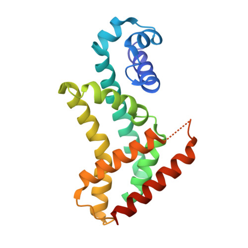
3PM1

Structure of QacR E90Q bound to Ethidium


Experimental Data Snapshot

  • Method: X-RAY DIFFRACTION
  • Resolution: 2.80 Å
  • R-Value Free: 0.280 
  • R-Value Work: 0.229 
  • R-Value Observed: 0.229 

Starting Model: experimental
View more details

wwPDB Validation   3D Report Full Report


Ligand Structure Quality Assessment 


This is version 1.2 of the entry. See complete history


Literature

A single acidic residue can guide binding site selection but does not govern QacR cationic-drug affinity.

Peters, K.M.Brooks, B.E.Schumacher, M.A.Skurray, R.A.Brennan, R.G.Brown, M.H.

(2011) PLoS One 6: e15974-e15974

  • DOI: https://doi.org/10.1371/journal.pone.0015974
  • Primary Citation of Related Structures:  
    3PM1

  • PubMed Abstract: 

    Structures of the multidrug-binding repressor protein QacR with monovalent and bivalent cationic drugs revealed that the carboxylate side-chains of E90 and E120 were proximal to the positively charged nitrogens of the ligands ethidium, malachite green and rhodamine 6G, and therefore may contribute to drug neutralization and binding affinity. Here, we report structural, biochemical and in vivo effects of substituting these glutamate residues. Unexpectedly, substitutions had little impact on ligand affinity or in vivo induction capabilities. Structures of QacR(E90Q) and QacR(E120Q) with ethidium or malachite green took similar global conformations that differed significantly from all previously described QacR-drug complexes but still prohibited binding to cognate DNA. Strikingly, the QacR(E90Q)-rhodamine 6G complex revealed two mutually exclusive rhodamine 6G binding sites. Despite multiple structural changes, all drug binding was essentially isoenergetic. Thus, these data strongly suggest that rather than contributing significantly to ligand binding affinity, the role of acidic residues lining the QacR multidrug-binding pocket is primarily to attract and guide cationic drugs to the "best available" positions within the pocket that elicit QacR induction.


  • Organizational Affiliation

    School of Biological Sciences, University of Sydney, Sydney, New South Wales, Australia.


Macromolecules
Find similar proteins by:  (by identity cutoff)  |  3D Structure
Entity ID: 1
MoleculeChains Sequence LengthOrganismDetailsImage
HTH-type transcriptional regulator qacRA [auth B],
B [auth A]
194Staphylococcus aureus subsp. aureus Mu50Mutation(s): 4 
Gene Names: qacR
UniProt
Find proteins for P0A0N3 (Staphylococcus aureus (strain Mu50 / ATCC 700699))
Explore P0A0N3 
Go to UniProtKB:  P0A0N3
Entity Groups  
Sequence Clusters30% Identity50% Identity70% Identity90% Identity95% Identity100% Identity
UniProt GroupP0A0N3
Sequence Annotations
Expand
  • Reference Sequence
Small Molecules
Ligands 2 Unique
IDChains Name / Formula / InChI Key2D Diagram3D Interactions
ET
Query on ET

Download Ideal Coordinates CCD File 
G [auth A]ETHIDIUM
C21 H20 N3
QTANTQQOYSUMLC-UHFFFAOYSA-O
SO4
Query on SO4

Download Ideal Coordinates CCD File 
C [auth B]
D [auth B]
E [auth B]
F [auth B]
H [auth A]
C [auth B],
D [auth B],
E [auth B],
F [auth B],
H [auth A],
I [auth A],
J [auth A]
SULFATE ION
O4 S
QAOWNCQODCNURD-UHFFFAOYSA-L
Binding Affinity Annotations 
IDSourceBinding Affinity
ET PDBBind:  3PM1 Kd: 1180 (nM) from 1 assay(s)
Experimental Data & Validation

Experimental Data

  • Method: X-RAY DIFFRACTION
  • Resolution: 2.80 Å
  • R-Value Free: 0.280 
  • R-Value Work: 0.229 
  • R-Value Observed: 0.229 
  • Space Group: P 62
Unit Cell:
Length ( Å )Angle ( ˚ )
a = 104.148α = 90
b = 104.148β = 90
c = 98.564γ = 120
Software Package:
Software NamePurpose
ADSCdata collection
MOLREPphasing
CNSrefinement
MOSFLMdata reduction
SCALAdata scaling

Structure Validation

View Full Validation Report


Ligand Structure Quality Assessment 


Entry History 

Deposition Data

Revision History  (Full details and data files)

  • Version 1.0: 2011-07-13
    Type: Initial release
  • Version 1.1: 2024-02-21
    Changes: Data collection, Database references, Derived calculations
  • Version 1.2: 2024-04-03
    Changes: Refinement description