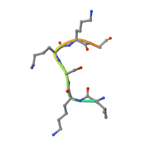
3S7F

Structural Basis of Substrate Methylation and Inhibition of SMYD2


Experimental Data Snapshot

  • Method: X-RAY DIFFRACTION
  • Resolution: 2.85 Å
  • R-Value Free: 0.201 
  • R-Value Work: 0.172 
  • R-Value Observed: 0.173 

wwPDB Validation   3D Report Full Report


Ligand Structure Quality Assessment 


This is version 1.2 of the entry. See complete history


Literature

Structural Basis of Substrate Methylation and Inhibition of SMYD2.

Ferguson, A.D.Larsen, N.A.Howard, T.Pollard, H.Green, I.Grande, C.Cheung, T.Garcia-Arenas, R.Cowen, S.Wu, J.Godin, R.Chen, H.Keen, N.

(2011) Structure 19: 1262-1273

  • DOI: https://doi.org/10.1016/j.str.2011.06.011
  • Primary Citation of Related Structures:  
    3S7B, 3S7D, 3S7F, 3S7J

  • PubMed Abstract: 

    Protein lysine methyltransferases are important regulators of epigenetic signaling. These enzymes catalyze the transfer of donor methyl groups from S-adenosylmethionine to specific acceptor lysines on histones, leading to changes in chromatin structure and transcriptional regulation. These enzymes also methylate nonhistone protein substrates, revealing an additional mechanism to regulate cellular physiology. The oncogenic protein SMYD2 represses the functional activities of the tumor suppressor proteins p53 and Rb, making it an attractive drug target. Here we report the discovery of AZ505, a potent and selective inhibitor of SMYD2 that was identified from a high throughput chemical screen. We also present the crystal structures of SMYD2 with p53 substrate and product peptides, and notably, in complex with AZ505. This substrate competitive inhibitor is bound in the peptide binding groove of SMYD2. These results have implications for the development of SMYD2 inhibitors, and indicate the potential for developing novel therapies targeting this target class.


  • Organizational Affiliation

    DECS Structural Chemistry, AstraZeneca, Waltham, MA 02451, USA. andrew.ferguson@astrazeneca.com


Macromolecules
Find similar proteins by:  (by identity cutoff)  |  3D Structure
Entity ID: 1
MoleculeChains Sequence LengthOrganismDetailsImage
N-lysine methyltransferase SMYD2433Homo sapiensMutation(s): 0 
Gene Names: SMYD2KMT3C
EC: 2.1.1 (PDB Primary Data), 2.1.1.43 (PDB Primary Data), 2.1.1.354 (UniProt)
UniProt & NIH Common Fund Data Resources
Find proteins for Q9NRG4 (Homo sapiens)
Explore Q9NRG4 
Go to UniProtKB:  Q9NRG4
PHAROS:  Q9NRG4
GTEx:  ENSG00000143499 
Entity Groups  
Sequence Clusters30% Identity50% Identity70% Identity90% Identity95% Identity100% Identity
UniProt GroupQ9NRG4
Sequence Annotations
Expand
  • Reference Sequence

Find similar proteins by:  Sequence   |   3D Structure  

Entity ID: 2
MoleculeChains Sequence LengthOrganismDetailsImage
p53 peptideB [auth I]13N/AMutation(s): 0 
Entity Groups  
Sequence Clusters30% Identity50% Identity70% Identity90% Identity95% Identity100% Identity
Sequence Annotations
Expand
  • Reference Sequence
Experimental Data & Validation

Experimental Data

  • Method: X-RAY DIFFRACTION
  • Resolution: 2.85 Å
  • R-Value Free: 0.201 
  • R-Value Work: 0.172 
  • R-Value Observed: 0.173 
  • Space Group: I 4
Unit Cell:
Length ( Å )Angle ( ˚ )
a = 156.413α = 90
b = 156.413β = 90
c = 52.895γ = 90
Software Package:
Software NamePurpose
ADSCdata collection
MOLREPphasing
BUSTERrefinement
XDSdata reduction
SCALAdata scaling

Structure Validation

View Full Validation Report


Ligand Structure Quality Assessment 


Entry History 

Deposition Data

Revision History  (Full details and data files)

  • Version 1.0: 2011-08-10
    Type: Initial release
  • Version 1.1: 2011-09-21
    Changes: Database references
  • Version 1.2: 2024-02-28
    Changes: Data collection, Database references, Derived calculations