3SN2 | pdb_00003sn2

Crystal structure analysis of iron regulatory protein 1 in complex with transferrin receptor IRE B RNA


Experimental Data Snapshot

  • Method: X-RAY DIFFRACTION
  • Resolution: 2.99 Å
  • R-Value Free: 
    0.275 (Depositor), 0.274 (DCC) 
  • R-Value Work: 
    0.227 (Depositor), 0.226 (DCC) 
  • R-Value Observed: 
    0.230 (Depositor) 

Starting Model: experimental
View more details

wwPDB Validation 3D Report Full Report

Validation slider image for 3SN2

This is version 1.3 of the entry. See complete history

Literature

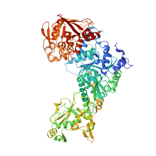
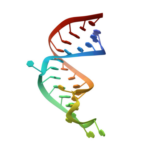
Accommodating variety in iron-responsive elements: Crystal structure of transferrin receptor 1 B IRE bound to iron regulatory protein 1.

Walden, W.E.Selezneva, A.Volz, K.

(2012) FEBS Lett 586: 32-35

  • DOI: https://doi.org/10.1016/j.febslet.2011.11.018
  • Primary Citation Related Structures: 
    3SN2

  • PubMed Abstract: 

    Iron responsive elements (IREs) are short stem-loop structures found in several mRNAs encoding proteins involved in cellular iron metabolism. Iron regulatory proteins (IRPs) control iron homeostasis through differential binding to the IREs, accommodating any sequence or structural variations that the IREs may present. Here we report the structure of IRP1 in complex with transferrin receptor 1 B (TfR B) IRE, and compare it to the complex with ferritin H (Ftn H) IRE. The two IREs are bound to IRP1 through nearly identical protein-RNA contacts, although their stem conformations are significantly different. These results support the view that binding of different IREs with IRP1 depends both on protein and RNA conformational plasticity, adapting to RNA variation while retaining conserved protein-RNA contacts.


  • Organizational Affiliation
    • Department of Microbiology and Immunology, University of Illinois at Chicago, Chicago, IL 60612, USA.

Macromolecule Content 

  • Total Structure Weight: 110.02 kDa 
  • Atom Count: 7,389 
  • Modeled Residue Count: 877 
  • Deposited Residue Count: 937 
  • Unique protein chains: 1
  • Unique nucleic acid chains: 1

Macromolecules


Find similar proteins by:|  3D Structure
Entity ID: 1
MoleculeChains  Sequence LengthOrganismDetailsImage
Cytoplasmic aconitate hydratase908Oryctolagus cuniculusMutation(s): 2 
Gene Names: ACO1FRPIREB1IREBPIRP1
EC: 4.2.1.3
UniProt
Find proteins for Q01059 (Oryctolagus cuniculus)
Explore Q01059 
Go to UniProtKB:  Q01059
Entity Groups
Sequence Clusters30% Identity50% Identity70% Identity90% Identity95% Identity100% Identity
UniProt GroupQ01059
Sequence Annotations
Expand
Reference Sequence
Find similar nucleic acids by:  (by identity cutoff) 
Entity ID: 2
MoleculeChains LengthOrganismImage
transferrin receptor iron regulatory element B RNA29N/A
Sequence Annotations
Expand
Reference Sequence

Experimental Data & Validation

Experimental Data

  • Method: X-RAY DIFFRACTION
  • Resolution: 2.99 Å
  • R-Value Free:  0.275 (Depositor), 0.274 (DCC) 
  • R-Value Work:  0.227 (Depositor), 0.226 (DCC) 
  • R-Value Observed: 0.230 (Depositor) 
Space Group: P 41 21 2
Unit Cell:
Length ( Å )Angle ( ˚ )
a = 101.675α = 90
b = 101.675β = 90
c = 281.349γ = 90
Software Package:
Software NamePurpose
SERGUIdata collection
PHASERphasing
PHENIXrefinement
X-GENdata reduction
X-GENdata scaling

Structure Validation

View Full Validation Report


Entry History 

Deposition Data

Revision History  (Full details and data files)

  • Version 1.0: 2011-11-23
    Type: Initial release
  • Version 1.1: 2014-12-24
    Changes: Database references
  • Version 1.2: 2017-11-08
    Changes: Refinement description
  • Version 1.3: 2023-09-13
    Changes: Data collection, Database references, Refinement description