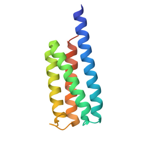
3U3B | pdb_00003u3b

Crystal Structure of Computationally Redesigned Four-Helix Bundle


Experimental Data Snapshot

  • Method: X-RAY DIFFRACTION
  • Resolution: 1.85 Å
  • R-Value Free: 
    0.214 (Depositor), 0.203 (DCC) 
  • R-Value Work: 
    0.176 (Depositor) 
  • R-Value Observed: 
    0.178 (Depositor) 

Starting Model: in silico
View more details

wwPDB Validation 3D Report Full Report

Validation slider image for 3U3B

This is version 1.4 of the entry. See complete history

Literature

Increasing sequence diversity with flexible backbone protein design: the complete redesign of a protein hydrophobic core.

Murphy, G.S.Mills, J.L.Miley, M.J.Machius, M.Szyperski, T.Kuhlman, B.

(2012) Structure 20: 1086-1096

  • DOI: https://doi.org/10.1016/j.str.2012.03.026
  • Primary Citation Related Structures: 
    3U3B

  • PubMed Abstract: 

    Protein design tests our understanding of protein stability and structure. Successful design methods should allow the exploration of sequence space not found in nature. However, when redesigning naturally occurring protein structures, most fixed backbone design algorithms return amino acid sequences that share strong sequence identity with wild-type sequences, especially in the protein core. This behavior places a restriction on functional space that can be explored and is not consistent with observations from nature, where sequences of low identity have similar structures. Here, we allow backbone flexibility during design to mutate every position in the core (38 residues) of a four-helix bundle protein. Only small perturbations to the backbone, 1-2 Å, were needed to entirely mutate the core. The redesigned protein, DRNN, is exceptionally stable (melting point >140°C). An NMR and X-ray crystal structure show that the side chains and backbone were accurately modeled (all-atom RMSD = 1.3 Å).


  • Organizational Affiliation
    • Department of Chemistry, University of North Carolina at Chapel Hill, Chapel Hill, NC 27599-3290, USA.

Macromolecule Content 

  • Total Structure Weight: 26.53 kDa 
  • Atom Count: 1,875 
  • Modeled Residue Count: 206 
  • Deposited Residue Count: 226 
  • Unique protein chains: 1

Macromolecules

Find similar proteins by:|  3D Structure
Entity ID: 1
MoleculeChains  Sequence LengthOrganismDetailsImage
computationally designed four-helix bundle protein
A, B
113synthetic constructMutation(s): 0 

Experimental Data & Validation

Experimental Data

  • Method: X-RAY DIFFRACTION
  • Resolution: 1.85 Å
  • R-Value Free:  0.214 (Depositor), 0.203 (DCC) 
  • R-Value Work:  0.176 (Depositor) 
  • R-Value Observed: 0.178 (Depositor) 
Space Group: P 1
Unit Cell:
Length ( Å )Angle ( ˚ )
a = 27.564α = 63.89
b = 43.958β = 80.03
c = 47.68γ = 87.02
Software Package:
Software NamePurpose
PHASERphasing
PHENIXrefinement
HKL-3000data reduction
HKL-3000data scaling

Structure Validation

View Full Validation Report


Entry History 

Deposition Data

Revision History  (Full details and data files)

  • Version 1.0: 2012-06-13
    Type: Initial release
  • Version 1.1: 2013-01-09
    Changes: Database references
  • Version 1.2: 2017-11-08
    Changes: Refinement description
  • Version 1.3: 2024-02-28
    Changes: Data collection, Database references
  • Version 1.4: 2024-04-03
    Changes: Refinement description