3UBP

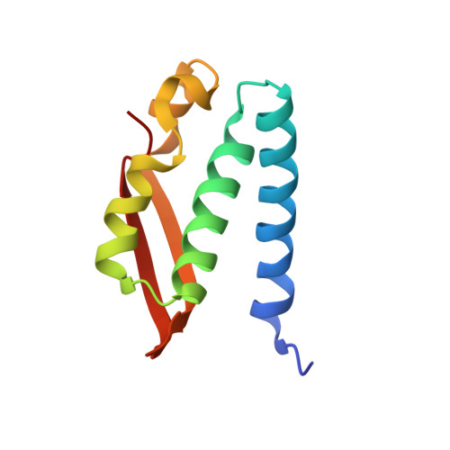
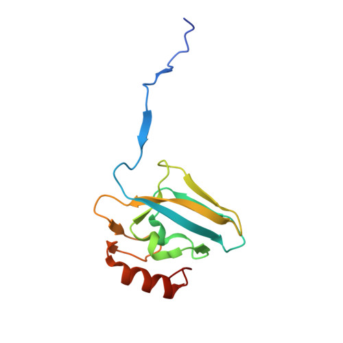
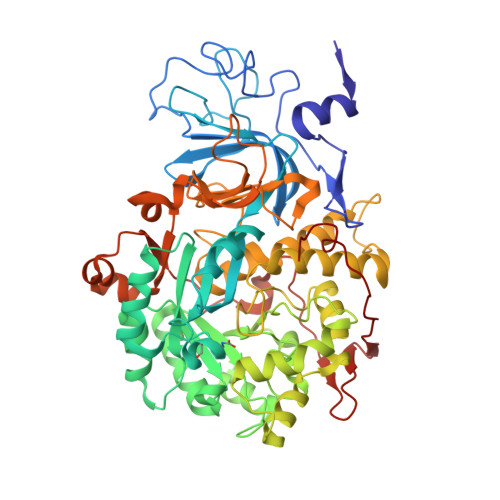
DIAMIDOPHOSPHATE INHIBITED BACILLUS PASTEURII UREASE


Experimental Data Snapshot

  • Method: X-RAY DIFFRACTION
  • Resolution: 2.00 Å
  • R-Value Free: 0.200 
  • R-Value Work: 0.158 
  • R-Value Observed: 0.158 

Starting Model: experimental
View more details

wwPDB Validation   3D Report Full Report


This is version 2.2 of the entry. See complete history


Literature

A new proposal for urease mechanism based on the crystal structures of the native and inhibited enzyme from Bacillus pasteurii: why urea hydrolysis costs two nickels.

Benini, S.Rypniewski, W.R.Wilson, K.S.Miletti, S.Ciurli, S.Mangani, S.

(1999) Structure 7: 205-216

  • DOI: https://doi.org/10.1016/S0969-2126(99)80026-4
  • Primary Citation of Related Structures:  
    2UBP, 3UBP

  • PubMed Abstract: 

    Urease catalyzes the hydrolysis of urea, the final step of organic nitrogen mineralization, using a bimetallic nickel centre. The role of the active site metal ions and amino acid residues has not been elucidated to date. Many pathologies are associated with the activity of ureolytic bacteria, and the efficiency of soil nitrogen fertilization with urea is severely decreased by urease activity. Therefore, the development of urease inhibitors would lead to a reduction of environmental pollution, to enhanced efficiency of nitrogen uptake by plants, and to improved therapeutic strategies for treatment of infections due to ureolytic bacteria. Structure-based design of urease inhibitors would require knowledge of the enzyme mechanism at the molecular level. The structures of native and inhibited urease from Bacillus pasteurii have been determined at a resolution of 2.0 A by synchrotron X-ray cryogenic crystallography. In the native enzyme, the coordination sphere of each of the two nickel ions is completed by a water molecule and a bridging hydroxide. A fourth water molecule completes a tetrahedral cluster of solvent molecules. The enzyme crystallized in the presence of phenylphosphorodiamidate contains the tetrahedral transition-state analogue diamidophosphoric acid, bound to the two nickel ions in an unprecedented mode. Comparison of the native and inhibited structures reveals two distinct conformations of the flap lining the active-site cavity. The mode of binding of the inhibitor, and a comparison between the native and inhibited urease structures, indicate a novel mechanism for enzymatic urea hydrolysis which reconciles the available structural and biochemical data.


  • Organizational Affiliation

    European Molecular Biology Laboratory, DESY, Notkestrasse 85 D-22603, Hamburg, Germany.


Macromolecules
Find similar proteins by:  (by identity cutoff)  |  3D Structure
Entity ID: 1
MoleculeChains Sequence LengthOrganismDetailsImage
PROTEIN (UREASE GAMMA SUBUNIT)101Sporosarcina pasteuriiMutation(s): 0 
EC: 3.5.1.5
UniProt
Find proteins for P41022 (Sporosarcina pasteurii)
Go to UniProtKB:  P41022
Sequence Annotations
Expand
  • Reference Sequence
Find similar proteins by:  (by identity cutoff)  |  3D Structure
Entity ID: 2
MoleculeChains Sequence LengthOrganismDetailsImage
PROTEIN (UREASE BETA SUBUNIT)126Sporosarcina pasteuriiMutation(s): 0 
EC: 3.5.1.5
UniProt
Find proteins for P41021 (Sporosarcina pasteurii)
Explore P41021 
Go to UniProtKB:  P41021
Entity Groups  
Sequence Clusters30% Identity50% Identity70% Identity90% Identity95% Identity100% Identity
UniProt GroupP41021
Sequence Annotations
Expand
  • Reference Sequence
Find similar proteins by:  (by identity cutoff)  |  3D Structure
Entity ID: 3
MoleculeChains Sequence LengthOrganismDetailsImage
PROTEIN (UREASE ALPHA SUBUNIT)570Sporosarcina pasteuriiMutation(s): 7 
EC: 3.5.1.5
UniProt
Find proteins for P41020 (Sporosarcina pasteurii)
Explore P41020 
Go to UniProtKB:  P41020
Entity Groups  
Sequence Clusters30% Identity50% Identity70% Identity90% Identity95% Identity100% Identity
UniProt GroupP41020
Sequence Annotations
Expand
  • Reference Sequence
Small Molecules
Modified Residues  1 Unique
IDChains TypeFormula2D DiagramParent
KCX
Query on KCX
C
L-PEPTIDE LINKINGC7 H14 N2 O4LYS
Experimental Data & Validation

Experimental Data

  • Method: X-RAY DIFFRACTION
  • Resolution: 2.00 Å
  • R-Value Free: 0.200 
  • R-Value Work: 0.158 
  • R-Value Observed: 0.158 
  • Space Group: P 63 2 2
Unit Cell:
Length ( Å )Angle ( ˚ )
a = 131.532α = 90
b = 131.532β = 90
c = 188.451γ = 120
Software Package:
Software NamePurpose
AMoREphasing
REFMACrefinement
DENZOdata reduction
SCALEPACKdata scaling

Structure Validation

View Full Validation Report


Entry History 

Deposition Data

Revision History  (Full details and data files)

  • Version 1.0: 1999-12-17
    Type: Initial release
  • Version 1.1: 2007-10-16
    Changes: Version format compliance
  • Version 1.2: 2011-07-13
    Changes: Derived calculations, Version format compliance
  • Version 2.0: 2023-05-31
    Changes: Atomic model, Data collection, Database references, Derived calculations
  • Version 2.1: 2023-09-20
    Changes: Advisory, Data collection, Derived calculations, Refinement description
  • Version 2.2: 2023-11-15
    Changes: Data collection