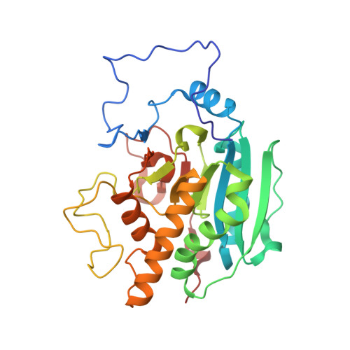
3V0P

Crystal structure of the Fucosylgalactoside alpha N-acetylgalactosaminyltransferase (GTA, cisAB mutant L266G, G268A) in complex with a novel UDP-Gal derived inhibitor (4GW) and H-antigen acceptor


Experimental Data Snapshot

  • Method: X-RAY DIFFRACTION
  • Resolution: 1.90 Å
  • R-Value Free: 0.193 
  • R-Value Work: 0.159 
  • R-Value Observed: 0.161 

Starting Model: experimental
View more details

wwPDB Validation   3D Report Full Report


Ligand Structure Quality Assessment 


This is version 1.5 of the entry. See complete history


Literature

Base-modified Donor Analogues Reveal Novel Dynamic Features of a Glycosyltransferase.

Jrgensen, R.Pesnot, T.Lee, H.J.Palcic, M.M.Wagner, G.K.

(2013) J Biol Chem 288: 26201-26208

  • DOI: https://doi.org/10.1074/jbc.M113.465963
  • Primary Citation of Related Structures:  
    3V0L, 3V0M, 3V0N, 3V0O, 3V0P, 3V0Q

  • PubMed Abstract: 

    Glycosyltransferases (GTs) are enzymes that are involved, as Nature's "glycosylation reagents," in many fundamental biological processes including cell adhesion and blood group biosynthesis. Although of similar importance to that of other large enzyme families such as protein kinases and proteases, the undisputed potential of GTs for chemical biology and drug discovery has remained largely unrealized to date. This is due, at least in part, to a relative lack of GT inhibitors and tool compounds for structural, mechanistic, and cellular studies. In this study, we have used a novel class of GT donor analogues to obtain new structural and enzymological information for a representative blood group GT. These analogues interfere with the folding of an internal loop and the C terminus, which are essential for catalysis. Our experiments have led to the discovery of an entirely new active site folding mode for this enzyme family, which can be targeted in inhibitor development, similar to the DFG motif in protein kinases. Taken together, our results provide new insights into substrate binding, dynamics, and utilization in this important enzyme family, which can very likely be harnessed for the rational development of new GT inhibitors and probes.


  • Organizational Affiliation

    From the Department of Microbiology and Infection Control, Statens Serum Institut, DK-2300 Copenhagen S, Denmark,; the Carlsberg Laboratory, Gamle Carlsberg Vej 10, DK-1799 Copenhagen V, Denmark,. Electronic address: renj@ssi.dk.


Macromolecules
Find similar proteins by:  (by identity cutoff)  |  3D Structure
Entity ID: 1
MoleculeChains Sequence LengthOrganismDetailsImage
Histo-blood group ABO system transferase
A, B
298Homo sapiensMutation(s): 2 
Gene Names: AB0ABO
EC: 2.4.1.40 (PDB Primary Data), 2.4.1.37 (PDB Primary Data)
UniProt & NIH Common Fund Data Resources
Find proteins for P16442 (Homo sapiens)
Explore P16442 
Go to UniProtKB:  P16442
PHAROS:  P16442
Entity Groups  
Sequence Clusters30% Identity50% Identity70% Identity90% Identity95% Identity100% Identity
UniProt GroupP16442
Sequence Annotations
Expand
  • Reference Sequence
Small Molecules
Ligands 4 Unique
IDChains Name / Formula / InChI Key2D Diagram3D Interactions
4GW
Query on 4GW

Download Ideal Coordinates CCD File 
D [auth A],
H [auth B]
5-(5-formylthiophen-2-yl)uridine 5'-(trihydrogen diphosphate)
C14 H16 N2 O13 P2 S
JAVHWQPALRUGBE-UORFTKCHSA-N
BHE
Query on BHE

Download Ideal Coordinates CCD File 
E [auth A],
I [auth B]
octyl 2-O-(6-deoxy-alpha-L-galactopyranosyl)-beta-D-galactopyranoside
C20 H38 O10
GTTDTLMUWQMDNA-ARNYJBIMSA-N
SO4
Query on SO4

Download Ideal Coordinates CCD File 
F [auth A]SULFATE ION
O4 S
QAOWNCQODCNURD-UHFFFAOYSA-L
MN
Query on MN

Download Ideal Coordinates CCD File 
C [auth A],
G [auth B]
MANGANESE (II) ION
Mn
WAEMQWOKJMHJLA-UHFFFAOYSA-N
Binding Affinity Annotations 
IDSourceBinding Affinity
4GW PDBBind:  3V0P Ki: 530 (nM) from 1 assay(s)
Experimental Data & Validation

Experimental Data

  • Method: X-RAY DIFFRACTION
  • Resolution: 1.90 Å
  • R-Value Free: 0.193 
  • R-Value Work: 0.159 
  • R-Value Observed: 0.161 
  • Space Group: P 21 21 2
Unit Cell:
Length ( Å )Angle ( ˚ )
a = 78.3α = 90
b = 153.52β = 90
c = 52.83γ = 90
Software Package:
Software NamePurpose
XSCALEdata scaling
PHASERphasing
PHENIXrefinement
PDB_EXTRACTdata extraction
MAR345data collection
XDSdata reduction

Structure Validation

View Full Validation Report


Ligand Structure Quality Assessment 


Entry History 

Deposition Data

Revision History  (Full details and data files)

  • Version 1.0: 2013-01-23
    Type: Initial release
  • Version 1.1: 2013-08-28
    Changes: Database references
  • Version 1.2: 2013-09-25
    Changes: Database references
  • Version 1.3: 2014-11-12
    Changes: Structure summary
  • Version 1.4: 2017-11-08
    Changes: Data collection, Refinement description
  • Version 1.5: 2023-09-13
    Changes: Data collection, Database references, Derived calculations, Refinement description