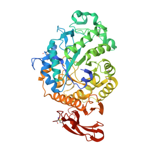
3VM7

Structure of an Alpha-Amylase from Malbranchea cinnamomea


Experimental Data Snapshot

  • Method: X-RAY DIFFRACTION
  • Resolution: 2.25 Å
  • R-Value Free: 0.191 
  • R-Value Work: 0.143 
  • R-Value Observed: 0.147 

wwPDB Validation   3D Report Full Report


Ligand Structure Quality Assessment 


This is version 1.2 of the entry. See complete history


Literature

A Novel Multifunctional alpha-Amylase from the Thermophilic Fungus Malbranchea cinnamomea: Biochemical Characterization and Three-Dimensional Structure.

Han, P.Zhou, P.Hu, S.Yang, S.Yan, Q.Jiang, Z.Q.

(2013) Appl Biochem Biotechnol 170: 420-435

  • DOI: https://doi.org/10.1007/s12010-013-0198-y
  • Primary Citation of Related Structures:  
    3VM7

  • PubMed Abstract: 

    A novel α-amylase (McAmyA) from the thermophilic fungus, Malbranchea cinnamomea was purified, characterized and crystallized in the present study. McAmyA was purified to apparent homogeneity with a molecular mass of 60.3 kDa on SDS-PAGE. The enzyme exhibited maximal activity at pH 6.5 and was stable within pH 5.0-10.0. It was most active at 65 °C and was stable up to 50 °C. McAmyA was capable of hydrolyzing amylose, starch, amylopectin, pullulan, cyclodextrins and maltooligosaccharides. The full-length cDNA of an α-amylase gene (McAmyA) from the strain was cloned. McAmyA consisted of a 1,476-bp open reading frame encoding 492 amino acids. It displayed the highest amino acid sequence homology (less than 60 %) with the reported α-amylases. The crystal structure of McAmyA was solved at a resolution of 2.25 Å (PDB code 3VM7). The overall structure of McAmyA reveals three domains with ten α helices and 14 β strands, and the putative catalytic residues are positioned at domain A with somewhat different secondary structural circumstances compared with typical α-amylases.


  • Organizational Affiliation

    Department of Biotechnology, College of Food Science and Nutritional Engineering, China Agricultural University, PO Box 294, No.17 Qinghua Donglu, Haidian District, Beijing, 100083, China.


Macromolecules
Find similar proteins by:  (by identity cutoff)  |  3D Structure
Entity ID: 1
MoleculeChains Sequence LengthOrganismDetailsImage
Alpha-amylase492Malbranchea cinnamomeaMutation(s): 0 
UniProt
Find proteins for K9L8F3 (Malbranchea cinnamomea)
Go to UniProtKB:  K9L8F3
Glycosylation
Glycosylation Sites: 1
Sequence Annotations
Expand
  • Reference Sequence
Small Molecules
Ligands 4 Unique
IDChains Name / Formula / InChI Key2D Diagram3D Interactions
NAG
Query on NAG

Download Ideal Coordinates CCD File 
C [auth A]2-acetamido-2-deoxy-beta-D-glucopyranose
C8 H15 N O6
OVRNDRQMDRJTHS-FMDGEEDCSA-N
GLC
Query on GLC

Download Ideal Coordinates CCD File 
D [auth A]alpha-D-glucopyranose
C6 H12 O6
WQZGKKKJIJFFOK-DVKNGEFBSA-N
TRS
Query on TRS

Download Ideal Coordinates CCD File 
E [auth A]2-AMINO-2-HYDROXYMETHYL-PROPANE-1,3-DIOL
C4 H12 N O3
LENZDBCJOHFCAS-UHFFFAOYSA-O
CA
Query on CA

Download Ideal Coordinates CCD File 
B [auth A]CALCIUM ION
Ca
BHPQYMZQTOCNFJ-UHFFFAOYSA-N
Experimental Data & Validation

Experimental Data

  • Method: X-RAY DIFFRACTION
  • Resolution: 2.25 Å
  • R-Value Free: 0.191 
  • R-Value Work: 0.143 
  • R-Value Observed: 0.147 
  • Space Group: P 1 21 1
Unit Cell:
Length ( Å )Angle ( ˚ )
a = 51.9α = 90
b = 84.94β = 105.61
c = 56.41γ = 90
Software Package:
Software NamePurpose
CrystalCleardata collection
PHENIXmodel building
REFMACrefinement
MOSFLMdata reduction
SCALAdata scaling
PHENIXphasing

Structure Validation

View Full Validation Report


Ligand Structure Quality Assessment 


Entry History 

Deposition Data

Revision History  (Full details and data files)

  • Version 1.0: 2013-05-29
    Type: Initial release
  • Version 1.1: 2013-06-05
    Changes: Database references
  • Version 1.2: 2020-07-29
    Type: Remediation
    Reason: Carbohydrate remediation
    Changes: Data collection, Derived calculations, Structure summary