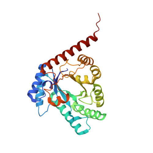
Structural insight into L-ribulose 3-epimerase from Mesorhizobium loti.
Uechi, K., Sakuraba, H., Yoshihara, A., Morimoto, K., Takata, G.(2013) Acta Crystallogr D Biol Crystallogr 69: 2330-2339
- PubMed: 24311575
- DOI: https://doi.org/10.1107/S0907444913021665
- Primary Citation of Related Structures:
3VYL - PubMed Abstract:
L-Ribulose 3-epimerase (L-RE) from Mesorhizobium loti has been identified as the first ketose 3-epimerase that shows the highest observed activity towards ketopentoses. In the present study, the crystal structure of the enzyme was determined to 2.7 Å resolution. The asymmetric unit contained two homotetramers with the monomer folded into an (α/β)8-barrel carrying four additional short α-helices. The overall structure of M. loti L-RE showed significant similarity to the structures of ketose 3-epimerases from Pseudomonas cichorii, Agrobacterium tumefaciens and Clostridium cellulolyticum, which use ketohexoses as preferred substrates. However, the size of the C-terminal helix (α8) was much larger in M. loti L-RE than the corresponding helices in the other enzymes. In M. loti L-RE the α8 helix and the following C-terminal tail possessed a unique subunit-subunit interface which promoted the formation of additional intermolecular interactions and strengthened the enzyme stability. Structural comparisons revealed that the relatively small hydrophobic pocket of the enzyme around the substrate was likely to be the main factor responsible for the marked specificity for ketopentoses shown by M. loti L-RE.
- Rare Sugar Research Center, Kagawa University, 2393 Ikenobe, Miki-cho, Kita-gun, Kagawa 761-0795, Japan.
Organizational Affiliation:


