RCSB PDB - 3ZXV: Crystal structure of Mycobacterium Tuberculosis Glutamine Synthetase in complex with tri-substituted imidazole inhibitor (4-(2-tert-butyl- 4-(6-methoxynaphthalen-2-yl)-1H-imidazol-5-yl)pyridin-2-amine) and L- methionine-S-sulfoximine phosphate

 3ZXV

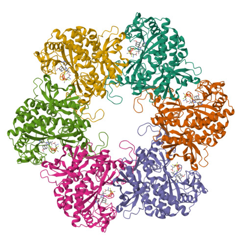
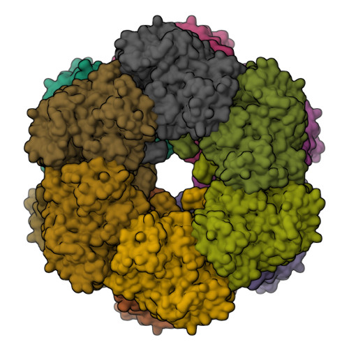
Crystal structure of Mycobacterium Tuberculosis Glutamine Synthetase in complex with tri-substituted imidazole inhibitor (4-(2-tert-butyl- 4-(6-methoxynaphthalen-2-yl)-1H-imidazol-5-yl)pyridin-2-amine) and L- methionine-S-sulfoximine phosphate


Experimental Data Snapshot

  • Method: X-RAY DIFFRACTION
  • Resolution: 2.26 Å
  • R-Value Free: 0.211 
  • R-Value Work: 0.195 
  • R-Value Observed: 0.196 

Starting Model: experimental
View more details

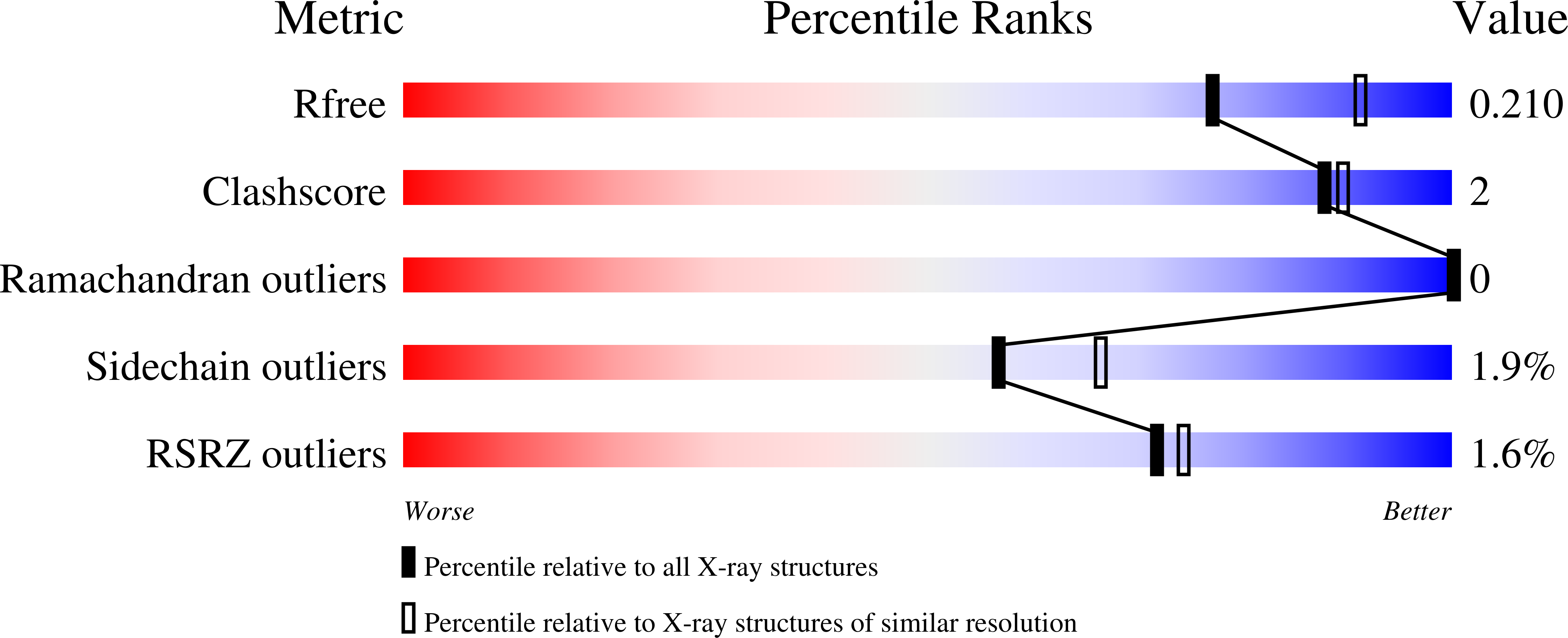
wwPDB Validation   3D Report Full Report


Ligand Structure Quality Assessment 

Created with Raphaël 2.3.0Worse 01 BetterLigand structure goodness of fit to experimental dataBest fitted MXIClick on this verticalbar to view detailsBest fitted P3SClick on this verticalbar to view details

This is version 1.3 of the entry. See complete history


Literature

Trisubstituted Imidazoles as Mycobacterium Tuberculosis Glutamine Synthetase Inhibitors.

Gising, J.Nilsson, M.T.Odell, L.R.Yahiaoui, S.Lindh, M.Iyer, H.Sinha, A.M.Srinivasa, B.R.Larhed, M.Mowbray, S.L.Karlen, A.

(2012) J Med Chem 55: 2894

  • DOI: https://doi.org/10.1021/jm201212h
  • Primary Citation of Related Structures:  
    3ZXR, 3ZXV

  • PubMed Abstract: 

    Mycobacterium tuberculosis glutamine synthetase (MtGS) is a promising target for antituberculosis drug discovery. In a recent high-throughput screening study we identified several classes of MtGS inhibitors targeting the ATP-binding site. We now explore one of these classes, the 2-tert-butyl-4,5-diarylimidazoles, and present the design, synthesis, and X-ray crystallographic studies leading to the identification of MtGS inhibitors with submicromolar IC(50) values and promising antituberculosis MIC values.


  • Organizational Affiliation

    Department of Medicinal Chemistry, Organic Pharmaceutical Chemistry, BMC, Uppsala University, Box 574, SE-751 23 Uppsala, Sweden.


Macromolecules
Find similar proteins by:  (by identity cutoff)  |  3D Structure
Entity ID: 1
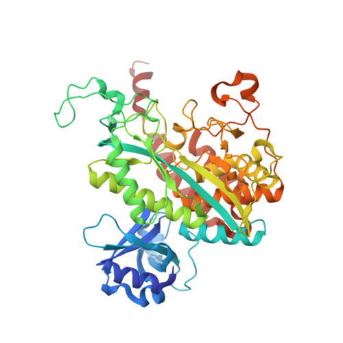
MoleculeChains Sequence LengthOrganismDetailsImage
GLUTAMINE SYNTHETASE 1
A, B, C, D, E
486Mycobacterium tuberculosis H37RvMutation(s): 0 
EC: 6.3.1.2
UniProt
Find proteins for P9WN39 (Mycobacterium tuberculosis (strain ATCC 25618 / H37Rv))
Explore P9WN39 
Go to UniProtKB:  P9WN39
Entity Groups  
Sequence Clusters30% Identity50% Identity70% Identity90% Identity95% Identity100% Identity
UniProt GroupP9WN39
Sequence Annotations
Expand
  • Reference Sequence
Small Molecules
Ligands 5 Unique
IDChains Name / Formula / InChI Key2D Diagram3D Interactions
MXI
Query on MXI

Download Ideal Coordinates CCD File 
BA [auth D]
G [auth A]
IA [auth E]
N [auth B]
PA [auth F]
4-(2-TERT-BUTYL-4-(6-METHOXYNAPHTHALEN-2-YL)-3H-IMIDAZOL-4-YL)PYRIDIN-2-AMINE
C23 H24 N4 O
KTHRIEHJGYTJED-UHFFFAOYSA-N
P3S
Query on P3S

Download Ideal Coordinates CCD File 
FA [auth D]
K [auth A]
MA [auth E]
R [auth B]
TA [auth F]
L-METHIONINE-S-SULFOXIMINE PHOSPHATE
C5 H13 N2 O6 P S
QQFOFBSCSWFFPB-NMAPHRJESA-N
PO4
Query on PO4

Download Ideal Coordinates CCD File 
GA [auth D]
L [auth A]
NA [auth E]
S [auth B]
UA [auth F]
PHOSPHATE ION
O4 P
NBIIXXVUZAFLBC-UHFFFAOYSA-K
CL
Query on CL

Download Ideal Coordinates CCD File 
AA [auth C]
HA [auth D]
M [auth A]
OA [auth E]
T [auth B]
CHLORIDE ION
Cl
VEXZGXHMUGYJMC-UHFFFAOYSA-M
MG
Query on MG

Download Ideal Coordinates CCD File 
CA [auth D]
DA [auth D]
EA [auth D]
H [auth A]
I [auth A]
MAGNESIUM ION
Mg
JLVVSXFLKOJNIY-UHFFFAOYSA-N
Binding Affinity Annotations 
IDSourceBinding Affinity
MXI PDBBind:  3ZXV IC50: 49 (nM) from 1 assay(s)
Experimental Data & Validation

Experimental Data

  • Method: X-RAY DIFFRACTION
  • Resolution: 2.26 Å
  • R-Value Free: 0.211 
  • R-Value Work: 0.195 
  • R-Value Observed: 0.196 
  • Space Group: C 2 2 21
Unit Cell:
Length ( Å )Angle ( ˚ )
a = 132.59α = 90
b = 227.15β = 90
c = 201.62γ = 90
Software Package:
Software NamePurpose
REFMACrefinement
XDSdata reduction
XSCALEdata scaling
REFMACphasing

Structure Validation

View Full Validation Report


Ligand Structure Quality Assessment 

Created with Raphaël 2.3.0Worse 01 BetterLigand structure goodness of fit to experimental dataBest fitted MXIClick on this verticalbar to view detailsBest fitted P3SClick on this verticalbar to view details

Entry History 

Deposition Data

Revision History  (Full details and data files)

  • Version 1.0: 2012-04-04
    Type: Initial release
  • Version 1.1: 2019-05-08
    Changes: Data collection, Experimental preparation, Other
  • Version 1.2: 2019-05-15
    Changes: Data collection, Experimental preparation
  • Version 1.3: 2023-12-20
    Changes: Data collection, Database references, Derived calculations, Other, Refinement description