4AII | pdb_00004aii

Crystal structure of the rat REM2 GTPase - G domain bound to GDP


Experimental Data Snapshot

  • Method: X-RAY DIFFRACTION
  • Resolution: 2.66 Å
  • R-Value Free: 
    0.324 (Depositor), 0.314 (DCC) 
  • R-Value Work: 
    0.243 (Depositor), 0.243 (DCC) 
  • R-Value Observed: 
    0.247 (Depositor) 

Starting Model: experimental
View more details

wwPDB Validation 3D Report Full Report

Validation slider image for 4AII

Ligand Structure Quality Assessment 


This is version 1.2 of the entry. See complete history

Literature

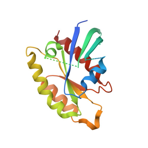
Structure of the Gdp-Bound G Domain of the Rgk Protein Rem2.

Reymond, P.Coquard, A.Chenon, M.Zeghouf, M.El Marjou, A.Thompson, A.Menetrey, J.

(2012) Acta Crystallogr Sect F Struct Biol Cryst Commun 68: 626

  • DOI: https://doi.org/10.1107/S1744309112013541
  • Primary Citation Related Structures: 
    4AII

  • PubMed Abstract: 

    RGK proteins are atypical small GTP-binding proteins that are involved in the regulation of voltage-dependent calcium channels and actin cytoskeleton remodelling. The structure of the Rem2 G domain bound to GDP is reported here in a monoclinic crystal form at 2.66 Å resolution. It is very similar to the structure determined previously from an orthorhombic crystal form. However, differences in the crystal-packing environment revealed that the switch I and switch II regions are flexible and not ordered as previously reported. Comparison of the available RGK protein structures along with those of other small GTP-binding proteins highlights two structural features characteristic of this atypical family and suggests that the conserved tryptophan residue in the DXWEX motif may be a structural determinant of the nucleotide-binding affinity.


  • Organizational Affiliation
    • Laboratoire d'Enzymologie et Biochimie Structurales, Centre de Recherche de Gif, CNRS, 91198 Gif-sur-Yvette, France.

Macromolecule Content 

  • Total Structure Weight: 41.24 kDa 
  • Atom Count: 2,258 
  • Modeled Residue Count: 290 
  • Deposited Residue Count: 360 
  • Unique protein chains: 1

Macromolecules

Find similar proteins by:|  3D Structure
Entity ID: 1
MoleculeChains  Sequence LengthOrganismDetailsImage
GTP-BINDING PROTEIN REM 2
A, B
180Rattus norvegicusMutation(s): 0 
UniProt
Find proteins for Q9WTY2 (Rattus norvegicus)
Explore Q9WTY2 
Go to UniProtKB:  Q9WTY2
Entity Groups
Sequence Clusters30% Identity50% Identity70% Identity90% Identity95% Identity100% Identity
UniProt GroupQ9WTY2
Sequence Annotations
Expand
Reference Sequence

Experimental Data & Validation

Experimental Data

  • Method: X-RAY DIFFRACTION
  • Resolution: 2.66 Å
  • R-Value Free:  0.324 (Depositor), 0.314 (DCC) 
  • R-Value Work:  0.243 (Depositor), 0.243 (DCC) 
  • R-Value Observed: 0.247 (Depositor) 
Space Group: P 1 21 1
Unit Cell:
Length ( Å )Angle ( ˚ )
a = 50.916α = 90
b = 59.374β = 101.51
c = 57.297γ = 90
Software Package:
Software NamePurpose
REFMACrefinement
XDSdata reduction
XDSdata scaling
PHASERphasing

Structure Validation

View Full Validation Report


Ligand Structure Quality Assessment 


Entry History 

Deposition Data

Revision History  (Full details and data files)

  • Version 1.0: 2012-06-13
    Type: Initial release
  • Version 1.1: 2012-06-20
    Changes: Other
  • Version 1.2: 2023-12-20
    Changes: Data collection, Database references, Derived calculations, Other, Refinement description