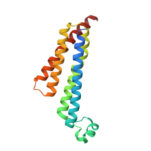
4AL1

Crystal structure of Human PS-1 GSH-analog complex


Experimental Data Snapshot

  • Method: X-RAY DIFFRACTION
  • Resolution: 1.95 Å
  • R-Value Free: 0.172 
  • R-Value Work: 0.172 
  • R-Value Observed: 0.163 

Starting Model: experimental
View more details

wwPDB Validation   3D Report Full Report


Ligand Structure Quality Assessment 


This is version 1.4 of the entry. See complete history


Literature

Crystal Structure of Microsomal Prostaglandin E2 Synthase Provides Insight Into Diversity in the Mapeg Superfamily.

Sjogren, T.Nord, J.Ek, M.Johansson, P.Liu, G.Geschwindner, S.

(2013) Proc Natl Acad Sci U S A 110: 3806

  • DOI: https://doi.org/10.1073/pnas.1218504110
  • Primary Citation of Related Structures:  
    4AL0, 4AL1

  • PubMed Abstract: 

    Prostaglandin E2 (PGE2) is a key mediator in inflammatory response. The main source of inducible PGE2, microsomal PGE2 synthase-1 (mPGES-1), has emerged as an interesting drug target for treatment of pain. To support inhibitor design, we have determined the crystal structure of human mPGES-1 to 1.2 Å resolution. The structure reveals three well-defined active site cavities within the membrane-spanning region in each monomer interface of the trimeric structure. An important determinant of the active site cavity is a small cytosolic domain inserted between transmembrane helices I and II. This extra domain is not observed in other structures of proteins within the MAPEG (Membrane-Associated Proteins involved in Eicosanoid and Glutathione metabolism) superfamily but is likely to be present also in microsomal GST-1 based on sequence similarity. An unexpected feature of the structure is a 16-Å-deep cone-shaped cavity extending from the cytosolic side into the membrane-spanning region. We suggest a potential role for this cavity in substrate access. Based on the structure of the active site, we propose a catalytic mechanism in which serine 127 plays a key role. We have also determined the structure of mPGES-1 in complex with a glutathione-based analog, providing insight into mPGES-1 flexibility and potential for structure-based drug design.


  • Organizational Affiliation

    Discovery Sciences, AstraZeneca R&D Mölndal, S-43183 Mölndal, Sweden. tove.sjogren@astrazeneca.com


Macromolecules
Find similar proteins by:  (by identity cutoff)  |  3D Structure
Entity ID: 1
MoleculeChains Sequence LengthOrganismDetailsImage
PROSTAGLANDIN E SYNTHASE152Homo sapiensMutation(s): 0 
EC: 5.3.99.3 (PDB Primary Data), 1.11.1 (UniProt), 2.5.1.18 (UniProt)
Membrane Entity: Yes 
UniProt & NIH Common Fund Data Resources
Find proteins for O14684 (Homo sapiens)
Explore O14684 
Go to UniProtKB:  O14684
PHAROS:  O14684
GTEx:  ENSG00000148344 
Entity Groups  
Sequence Clusters30% Identity50% Identity70% Identity90% Identity95% Identity100% Identity
UniProt GroupO14684
Sequence Annotations
Expand
  • Reference Sequence
Small Molecules
Ligands 4 Unique
IDChains Name / Formula / InChI Key2D Diagram3D Interactions
48T
Query on 48T

Download Ideal Coordinates CCD File 
F [auth A]L-gamma-glutamyl-S-(2-biphenyl-4-yl-2-oxoethyl)-L-cysteinylglycine
C24 H27 N3 O7 S
JIZFTRGOFNRYSA-OALUTQOASA-N
BOG
Query on BOG

Download Ideal Coordinates CCD File 
B [auth A]octyl beta-D-glucopyranoside
C14 H28 O6
HEGSGKPQLMEBJL-RKQHYHRCSA-N
PLM
Query on PLM

Download Ideal Coordinates CCD File 
C [auth A],
D [auth A]
PALMITIC ACID
C16 H32 O2
IPCSVZSSVZVIGE-UHFFFAOYSA-N
SO4
Query on SO4

Download Ideal Coordinates CCD File 
E [auth A]SULFATE ION
O4 S
QAOWNCQODCNURD-UHFFFAOYSA-L
Experimental Data & Validation

Experimental Data

  • Method: X-RAY DIFFRACTION
  • Resolution: 1.95 Å
  • R-Value Free: 0.172 
  • R-Value Work: 0.172 
  • R-Value Observed: 0.163 
  • Space Group: H 3
Unit Cell:
Length ( Å )Angle ( ˚ )
a = 76.623α = 90
b = 76.623β = 90
c = 123.349γ = 120
Software Package:
Software NamePurpose
REFMACrefinement
MOSFLMdata reduction
SCALAdata scaling
MOLREPphasing

Structure Validation

View Full Validation Report


Ligand Structure Quality Assessment 


Entry History 

Deposition Data

Revision History  (Full details and data files)

  • Version 1.0: 2013-02-06
    Type: Initial release
  • Version 1.1: 2013-02-27
    Changes: Database references
  • Version 1.2: 2013-04-03
    Changes: Database references
  • Version 1.3: 2020-07-29
    Type: Remediation
    Reason: Carbohydrate remediation
    Changes: Data collection, Derived calculations, Other, Structure summary
  • Version 1.4: 2023-12-20
    Changes: Data collection, Database references, Refinement description, Structure summary