4ASL | pdb_00004asl

Structure of Epa1A in complex with the T-antigen (Gal-b1-3- GalNAc)


Experimental Data Snapshot

  • Method: X-RAY DIFFRACTION
  • Resolution: 1.24 Å
  • R-Value Free: 
    0.155 (Depositor), 0.172 (DCC) 
  • R-Value Work: 
    0.117 (Depositor), 0.135 (DCC) 
  • R-Value Observed: 
    0.119 (Depositor) 

Starting Model: other
View more details

wwPDB Validation 3D Report Full Report

Validation slider image for 4ASL

This is version 3.2 of the entry. See complete history

Literature

Structural Basis for Promiscuity and Specificity During Candida Glabrata Invasion of Host Epithelia.

Maestre-Reyna, M.Diderrich, R.Veelders, M.S.Eulenburg, G.Kalugin, V.Bruckner, S.Keller, P.Rupp, S.Mosch, H.Essen, L.-O.

(2012) Proc Natl Acad Sci U S A 109: 16864

  • DOI: https://doi.org/10.1073/pnas.1207653109
  • Primary Citation Related Structures: 
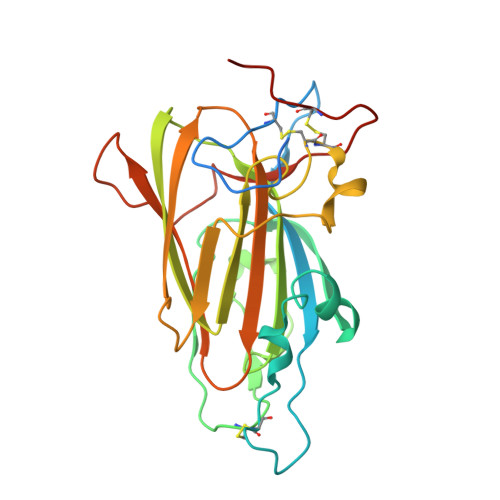
    4AF9, 4AFA, 4AFB, 4AFC, 4ASL

  • PubMed Abstract: 

    The human pathogenic yeast Candida glabrata harbors more than 20 surface-exposed, epithelial adhesins (Epas) for host cell adhesion. The Epa family recognizes host glycans and discriminates between target tissues by their adhesin (A) domains, but a detailed structural basis for ligand-binding specificity of Epa proteins has been lacking so far. In this study, we provide high-resolution crystal structures of the Epa1A domain in complex with different carbohydrate ligands that reveal how host cell mucin-type O-glycans are recognized and allow a structure-guided classification of the Epa family into specific subtypes. Further detailed structural and functional characterization of subtype-switched Epa1 variants shows that specificity is governed by two inner loops, CBL1 and CBL2, involved in calcium binding as well as by three outer loops, L1, L2, and L3. In summary, our study provides the structural basis for promiscuity and specificity of Epa adhesins, which might further contribute to developing anti-adhesive antimycotics and combating Candida colonization.


  • Organizational Affiliation
    • Biomedical Research Center/Department of Chemistry, Philipps-Universität Marburg, D-35032 Marburg, Germany.

Macromolecule Content 

  • Total Structure Weight: 29.88 kDa 
  • Atom Count: 2,484 
  • Modeled Residue Count: 229 
  • Deposited Residue Count: 259 
  • Unique protein chains: 1

Macromolecules

Find similar proteins by:|  3D Structure
Entity ID: 1
MoleculeChains  Sequence LengthOrganismDetailsImage
EPA1P259Nakaseomyces glabratusMutation(s): 0 
UniProt
Find proteins for Q6FUW5 (Candida glabrata (strain ATCC 2001 / BCRC 20586 / JCM 3761 / NBRC 0622 / NRRL Y-65 / CBS 138))
Explore Q6FUW5 
Go to UniProtKB:  Q6FUW5
Entity Groups
Sequence Clusters30% Identity50% Identity70% Identity90% Identity95% Identity100% Identity
UniProt GroupQ6FUW5
Sequence Annotations
Expand
Reference Sequence

Oligosaccharides

Help  
Entity ID: 2
MoleculeChains Length2D Diagram GlycosylationD Interactions
beta-D-galactopyranose-(1-3)-2-acetamido-2-deoxy-beta-D-galactopyranose
B
2N/A
Glycosylation Resources
GlyTouCan: G01534TU
GlyCosmos: G01534TU
GlyGen: G01534TU

Small Molecules

Ligands 3 Unique
IDChains Name / Formula / InChI Key2D Diagram3D Interactions
GOL

Query on GOL


Download:Ideal Coordinates CCD File
H [auth A],
I [auth A]
GLYCEROL
C3 H8 O3
PEDCQBHIVMGVHV-UHFFFAOYSA-N
CA

Query on CA


Download:Ideal Coordinates CCD File
C [auth A]CALCIUM ION
Ca
BHPQYMZQTOCNFJ-UHFFFAOYSA-N
NA

Query on NA


Download:Ideal Coordinates CCD File
D [auth A],
E [auth A],
F [auth A],
G [auth A],
J [auth A]
SODIUM ION
Na
FKNQFGJONOIPTF-UHFFFAOYSA-N

Experimental Data & Validation

Experimental Data

  • Method: X-RAY DIFFRACTION
  • Resolution: 1.24 Å
  • R-Value Free:  0.155 (Depositor), 0.172 (DCC) 
  • R-Value Work:  0.117 (Depositor), 0.135 (DCC) 
  • R-Value Observed: 0.119 (Depositor) 
Space Group: C 2 2 21
Unit Cell:
Length ( Å )Angle ( ˚ )
a = 74.6α = 89.9
b = 103.9β = 90
c = 69.4γ = 89.9
Software Package:
Software NamePurpose
REFMACrefinement
XDSdata reduction
SCALAdata scaling
XSCALEdata scaling
REFMACphasing

Structure Validation

View Full Validation Report


Entry History 

Deposition Data

Revision History  (Full details and data files)

  • Version 1.0: 2012-10-17
    Type: Initial release
  • Version 1.1: 2012-11-07
    Changes: Database references
  • Version 2.0: 2017-06-14
    Changes: Advisory, Atomic model, Derived calculations
  • Version 3.0: 2020-07-29
    Type: Remediation
    Reason: Carbohydrate remediation
    Changes: Advisory, Atomic model, Data collection, Derived calculations, Other, Structure summary
  • Version 3.1: 2024-05-01
    Changes: Data collection, Database references, Derived calculations, Refinement description, Structure summary
  • Version 3.2: 2024-11-06
    Changes: Structure summary