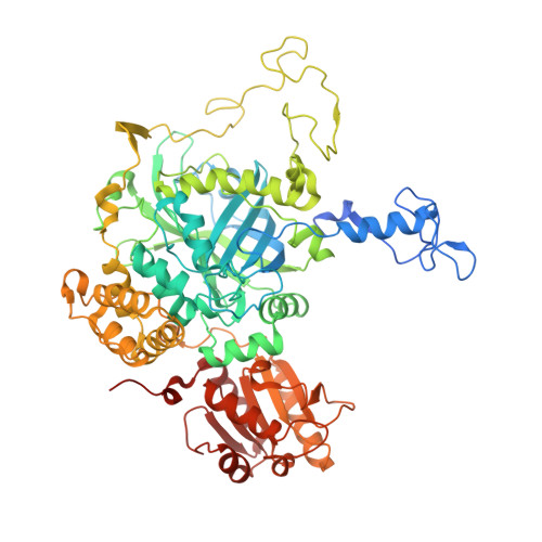
4AUL

Crystal structure, recombinant expression and mutagenesis studies of the bifunctional catalase-phenol oxidase from Scytalidium thermophilum


Experimental Data Snapshot

  • Method: X-RAY DIFFRACTION
  • Resolution: 1.50 Å
  • R-Value Free: 0.193 
  • R-Value Work: 0.143 
  • R-Value Observed: 0.145 

wwPDB Validation   3D Report Full Report


Ligand Structure Quality Assessment 


This is version 1.2 of the entry. See complete history


Literature

Structure, Recombinant Expression and Mutagenesis Studies of the Catalase with Oxidase Activity from Scytalidium Thermophilum

Yuzugullu, Y.Trinh, C.H.Smith, M.A.Pearson, A.R.Phillips, S.E.V.Sutay Kocabas, D.Bakir, U.Ogel, Z.B.McPherson, M.J.

(2013) Acta Crystallogr D Biol Crystallogr 69: 398

  • DOI: https://doi.org/10.1107/S0907444912049001
  • Primary Citation of Related Structures:  
    4AUE, 4AUL, 4AUM, 4AUN

  • PubMed Abstract: 

    Scytalidium thermophilum produces a catalase with phenol oxidase activity (CATPO) that catalyses the decomposition of hydrogen peroxide into oxygen and water and also oxidizes various phenolic compounds. A codon-optimized catpo gene was cloned and expressed in Escherichia coli. The crystal structures of native and recombinant S. thermophilum CATPO and two variants, H82N and V123F, were determined at resolutions of 2.7, 1.4, 1.5 and 1.9 Å, respectively. The structure of CATPO reveals a homotetramer with 698 residues per subunit and with strong structural similarity to Penicillium vitale catalase. The haem component is cis-hydroxychlorin γ-spirolactone, which is rotated 180° with respect to small-subunit catalases. The haem-binding pocket contains two highly conserved water molecules on the distal side. The H82N mutation resulted in conversion of the native d-type haem to a b-type haem. Kinetic studies of the H82N and V123F mutants indicate that both activities are likely to be associated with the haem centre and suggest that the secondary oxidase activity may be a general feature of catalases in the absence of hydrogen peroxide.


  • Organizational Affiliation

    Department of Biology, Kocaeli University, 41380 Kocaeli, Turkey.


Macromolecules
Find similar proteins by:  (by identity cutoff)  |  3D Structure
Entity ID: 1
MoleculeChains Sequence LengthOrganismDetailsImage
CATALASE-PHENOL OXIDASE
A, B, C, D
719Mycothermus thermophilusMutation(s): 0 
EC: 1.11.1.6
UniProt
Find proteins for M4GGR6 (Mycothermus thermophilus)
Go to UniProtKB:  M4GGR6
Sequence Annotations
Expand
  • Reference Sequence
Small Molecules
Ligands 2 Unique
IDChains Name / Formula / InChI Key2D Diagram3D Interactions
HEM
Query on HEM

Download Ideal Coordinates CCD File 
E [auth A],
I [auth B],
M [auth C],
Q [auth D]
PROTOPORPHYRIN IX CONTAINING FE
C34 H32 Fe N4 O4
KABFMIBPWCXCRK-RGGAHWMASA-L
CA
Query on CA

Download Ideal Coordinates CCD File 
F [auth A]
G [auth A]
H [auth A]
J [auth B]
K [auth B]
F [auth A],
G [auth A],
H [auth A],
J [auth B],
K [auth B],
L [auth B],
N [auth C],
O [auth C],
P [auth C],
R [auth D],
S [auth D],
T [auth D]
CALCIUM ION
Ca
BHPQYMZQTOCNFJ-UHFFFAOYSA-N
Experimental Data & Validation

Experimental Data

  • Method: X-RAY DIFFRACTION
  • Resolution: 1.50 Å
  • R-Value Free: 0.193 
  • R-Value Work: 0.143 
  • R-Value Observed: 0.145 
  • Space Group: C 1 2 1
Unit Cell:
Length ( Å )Angle ( ˚ )
a = 200.958α = 90
b = 121.919β = 115.28
c = 124.919γ = 90
Software Package:
Software NamePurpose
REFMACrefinement
XDSdata reduction
SCALAdata scaling

Structure Validation

View Full Validation Report


Ligand Structure Quality Assessment 


Entry History 

Deposition Data

Revision History  (Full details and data files)

  • Version 1.0: 2013-02-27
    Type: Initial release
  • Version 1.1: 2013-04-03
    Changes: Database references
  • Version 1.2: 2024-05-08
    Changes: Data collection, Database references, Derived calculations, Other