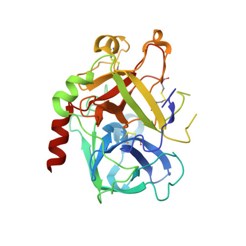
4BAQ

Thrombin in complex with inhibitor


Experimental Data Snapshot

  • Method: X-RAY DIFFRACTION
  • Resolution: 1.89 Å
  • R-Value Free: 0.221 
  • R-Value Work: 0.186 
  • R-Value Observed: 0.188 

wwPDB Validation   3D Report Full Report


Ligand Structure Quality Assessment 


This is version 1.5 of the entry. See complete history


Literature

Identification of Structure-Kinetic and Structure-Thermodynamic Relationships for Thrombin Inhibitors.

Winquist, J.Geschwindner, S.Xue, Y.Gustavsson, L.Musil, D.Deinum, J.Danielson, U.H.

(2013) Biochemistry 52: 613

  • DOI: https://doi.org/10.1021/bi301333z
  • Primary Citation of Related Structures:  
    4BAH, 4BAK, 4BAM, 4BAN, 4BAO, 4BAQ

  • PubMed Abstract: 

    To improve our understanding of drug-target interactions, we explored the effect of introducing substituted amine residues with increased chain length in the P3 residue of the thrombin inhibitor melagatran. Inhibition, kinetic, and thermodynamic data obtained via stopped-flow spectroscopy (SF), isothermal microcalorimetry (ITC), and surface plasmon resonance (SPR) biosensor analysis were interpreted with the help of X-ray crystal structures of the enzyme-inhibitor complexes. The association rate became faster when the lipophilicity of the inhibitors was increased. This was coupled to an increased enthalpic component and a corresponding decreased entropic component. The dissociation rates were reduced with an increase in chain length, with only a smaller increase and a decrease in the enthalpic and entropic components, respectively. Overall, the affinity increased with an increase in chain length, with similar changes in the enthalpic and entropic components. ITC analysis confirmed the equilibrium data from SPR analysis, showing that the interaction of melagatran was the most enthalpy-driven interaction. Structural analysis of the thrombin-inhibitor complex showed that the orientation of the P1 and P2 parts of the molecules was very similar, but that there were significant differences in the interaction between the terminal part of the P3 side chain and the binding pocket. A combination of charge repulsion, H-bonds, and hydrophobic interactions could be used to explain the observed kinetic and thermodynamic profiles for the ligands. In conclusion, changes in the structure of a lead compound can have significant effects on its interaction with the target that translate directly into kinetic and thermodynamic effects. In contrast to what may be intuitively expected, hydrogen bond formation and breakage are not necessarily reflected in enthalpy gains and losses, respectively.


  • Organizational Affiliation

    Department of Chemistry-BMC, Uppsala University, SE-751 23 Uppsala, Sweden.


Macromolecules
Find similar proteins by:  (by identity cutoff)  |  3D Structure
Entity ID: 1
MoleculeChains Sequence LengthOrganismDetailsImage
THROMBIN LIGHT CHAIN36Homo sapiensMutation(s): 0 
EC: 3.4.21.5
UniProt & NIH Common Fund Data Resources
Find proteins for P00734 (Homo sapiens)
Explore P00734 
Go to UniProtKB:  P00734
PHAROS:  P00734
GTEx:  ENSG00000180210 
Entity Groups  
Sequence Clusters30% Identity50% Identity70% Identity90% Identity95% Identity100% Identity
UniProt GroupP00734
Sequence Annotations
Expand
  • Reference Sequence
Find similar proteins by:  (by identity cutoff)  |  3D Structure
Entity ID: 2
MoleculeChains Sequence LengthOrganismDetailsImage
THROMBIN HEAVY CHAIN259Homo sapiensMutation(s): 0 
EC: 3.4.21.5
UniProt & NIH Common Fund Data Resources
Find proteins for P00734 (Homo sapiens)
Explore P00734 
Go to UniProtKB:  P00734
PHAROS:  P00734
GTEx:  ENSG00000180210 
Entity Groups  
Sequence Clusters30% Identity50% Identity70% Identity90% Identity95% Identity100% Identity
UniProt GroupP00734
Glycosylation
Glycosylation Sites: 1Go to GlyGen: P00734-1
Sequence Annotations
Expand
  • Reference Sequence

Find similar proteins by:  Sequence   |   3D Structure  

Entity ID: 3
MoleculeChains Sequence LengthOrganismDetailsImage
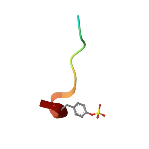
HIRUDIN VARIANT-1C [auth D]12Hirudo medicinalisMutation(s): 0 
UniProt
Find proteins for P01050 (Hirudo medicinalis)
Explore P01050 
Go to UniProtKB:  P01050
Entity Groups  
Sequence Clusters30% Identity50% Identity70% Identity90% Identity95% Identity100% Identity
UniProt GroupP01050
Sequence Annotations
Expand
  • Reference Sequence
Small Molecules
Modified Residues  1 Unique
IDChains TypeFormula2D DiagramParent
TYS
Query on TYS
C [auth D]L-PEPTIDE LINKINGC9 H11 N O6 STYR
Binding Affinity Annotations 
IDSourceBinding Affinity
M4Z PDBBind:  4BAQ Ki: 0.98 (nM) from 1 assay(s)
Experimental Data & Validation

Experimental Data

  • Method: X-RAY DIFFRACTION
  • Resolution: 1.89 Å
  • R-Value Free: 0.221 
  • R-Value Work: 0.186 
  • R-Value Observed: 0.188 
  • Space Group: C 1 2 1
Unit Cell:
Length ( Å )Angle ( ˚ )
a = 69.41α = 90
b = 71.51β = 100.34
c = 72.16γ = 90
Software Package:
Software NamePurpose
BUSTERrefinement
MOSFLMdata reduction
SCALAdata scaling

Structure Validation

View Full Validation Report


Ligand Structure Quality Assessment 


Entry History 

Deposition Data

Revision History  (Full details and data files)

  • Version 1.0: 2013-01-16
    Type: Initial release
  • Version 1.1: 2013-02-20
    Changes: Database references
  • Version 1.2: 2013-03-13
    Changes: Derived calculations
  • Version 1.3: 2017-07-05
    Changes: Data collection
  • Version 1.4: 2020-07-29
    Type: Remediation
    Reason: Carbohydrate remediation
    Changes: Data collection, Derived calculations, Other, Structure summary
  • Version 1.5: 2024-11-06
    Changes: Data collection, Database references, Structure summary