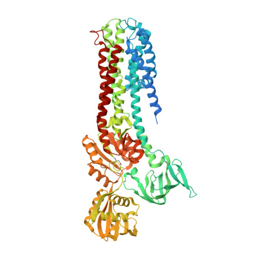
4BBJ

Copper-transporting PIB-ATPase in complex with beryllium fluoride representing the E2P state


Experimental Data Snapshot

  • Method: X-RAY DIFFRACTION
  • Resolution: 2.75 Å
  • R-Value Free: 0.247 
  • R-Value Work: 0.199 
  • R-Value Observed: 0.202 

Starting Model: experimental
View more details

wwPDB Validation   3D Report Full Report


Ligand Structure Quality Assessment 


This is version 1.3 of the entry. See complete history


Literature

Copper-Transporting P-Type Atpases Use a Unique Ion-Release Pathway

Andersson, M.Mattle, D.Sitsel, O.Klymchuk, T.Nielsen, A.Moller, L.B.White, S.H.Nissen, P.Gourdon, P.

(2014) Nat Struct Mol Biol 21: 43

  • DOI: https://doi.org/10.1038/nsmb.2721
  • Primary Citation of Related Structures:  
    4BBJ

  • PubMed Abstract: 

    Heavy metals in cells are typically regulated by PIB-type ATPases. The first structure of the class, a Cu(+)-ATPase from Legionella pneumophila (LpCopA), outlined a copper transport pathway across the membrane, which was inferred to be occluded. Here we show by molecular dynamics simulations that extracellular water solvated the transmembrane (TM) domain, results indicative of a Cu(+)-release pathway. Furthermore, a new LpCopA crystal structure determined at 2.8-Å resolution, trapped in the preceding E2P state, delineated the same passage, and site-directed-mutagenesis activity assays support a functional role for the conduit. The structural similarities between the TM domains of the two conformations suggest that Cu(+)-ATPases couple dephosphorylation and ion extrusion differently than do the well-characterized PII-type ATPases. The ion pathway explains why certain Menkes' and Wilson's disease mutations impair protein function and points to a site for inhibitors targeting pathogens.


  • Organizational Affiliation

    1] Department of Physiology and Biophysics, University of California at Irvine, Irvine, California, USA. [2] [3].


Macromolecules
Find similar proteins by:  (by identity cutoff)  |  3D Structure
Entity ID: 1
MoleculeChains Sequence LengthOrganismDetailsImage
COPPER EFFLUX ATPASE736Legionella pneumophila subsp. pneumophilaMutation(s): 0 
EC: 3.6.3 (PDB Primary Data), 7.2.2.8 (UniProt)
Membrane Entity: Yes 
UniProt
Find proteins for Q5ZWR1 (Legionella pneumophila subsp. pneumophila (strain Philadelphia 1 / ATCC 33152 / DSM 7513))
Explore Q5ZWR1 
Go to UniProtKB:  Q5ZWR1
Entity Groups  
Sequence Clusters30% Identity50% Identity70% Identity90% Identity95% Identity100% Identity
UniProt GroupQ5ZWR1
Sequence Annotations
Expand
  • Reference Sequence
Small Molecules
Modified Residues  1 Unique
IDChains TypeFormula2D DiagramParent
BFD
Query on BFD
A
L-PEPTIDE LINKINGC4 H6 Be F3 N O4ASP
Experimental Data & Validation

Experimental Data

  • Method: X-RAY DIFFRACTION
  • Resolution: 2.75 Å
  • R-Value Free: 0.247 
  • R-Value Work: 0.199 
  • R-Value Observed: 0.202 
  • Space Group: C 1 2 1
Unit Cell:
Length ( Å )Angle ( ˚ )
a = 242.01α = 90
b = 71.37β = 100.01
c = 72.43γ = 90
Software Package:
Software NamePurpose
PHENIXrefinement
XDSdata reduction
XSCALEdata scaling
PHASERphasing

Structure Validation

View Full Validation Report


Ligand Structure Quality Assessment 


Entry History 

Deposition Data

Revision History  (Full details and data files)

  • Version 1.0: 2013-12-11
    Type: Initial release
  • Version 1.1: 2015-04-15
    Changes: Structure summary
  • Version 1.2: 2023-12-20
    Changes: Data collection, Database references, Derived calculations, Other, Refinement description
  • Version 1.3: 2024-11-06
    Changes: Structure summary